 Movie
Movie Controller


+ Open data

- Basic information
| Entry | Database: PDB / ID: 7rcl | ||||||
|---|---|---|---|---|---|---|---|
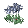
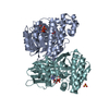
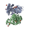
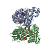
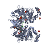
| Title | Crystal Structure of ADP-bound Galactokinase | ||||||
 Components Components | Galactokinase | ||||||
 Keywords Keywords | SUGAR BINDING PROTEIN / GALK / galactokinase / Galactose / ADP / disulfide | ||||||
| Function / homology |  Function and homology information Function and homology informationglycolytic process from galactose / Defective GALK1 causes GALCT2 / galactitol metabolic process / galactokinase / galactokinase activity / galactose catabolic process via UDP-galactose, Leloir pathway / galactose binding / galactose metabolic process / Galactose catabolism / extracellular exosome ...glycolytic process from galactose / Defective GALK1 causes GALCT2 / galactitol metabolic process / galactokinase / galactokinase activity / galactose catabolic process via UDP-galactose, Leloir pathway / galactose binding / galactose metabolic process / Galactose catabolism / extracellular exosome / ATP binding / membrane / cytoplasm / cytosol Similarity search - Function | ||||||
| Biological species |  Homo sapiens (human) Homo sapiens (human) | ||||||
| Method |  X-RAY DIFFRACTION / SYNCHROTRON / MOLECULAR REPLACEMENT / Resolution: 2.4 Å X-RAY DIFFRACTION / SYNCHROTRON / MOLECULAR REPLACEMENT / Resolution: 2.4 Å | ||||||
| Model details | Human Galactokinase enzyme with ADP bound | ||||||
 Authors Authors | Whitby, F.G. / Hall, M.D. | ||||||
| Funding support |  United States, 1items United States, 1items
| ||||||
 Citation Citation | Journal: J.Med.Chem. / Year: 2021 Title: Structure-Based Optimization of Small Molecule Human Galactokinase Inhibitors. Authors: Liu, L. / Tang, M. / Pragani, R. / Whitby, F.G. / Zhang, Y.Q. / Balakrishnan, B. / Fang, Y. / Karavadhi, S. / Tao, D. / LeClair, C.A. / Hall, M.D. / Marugan, J.J. / Boxer, M. / Shen, M. / ...Authors: Liu, L. / Tang, M. / Pragani, R. / Whitby, F.G. / Zhang, Y.Q. / Balakrishnan, B. / Fang, Y. / Karavadhi, S. / Tao, D. / LeClair, C.A. / Hall, M.D. / Marugan, J.J. / Boxer, M. / Shen, M. / Hill, C.P. / Lai, K. / Patnaik, S. | ||||||
| History |
|
- Structure visualization
| Structure viewer | Molecule: MolmilJmol/JSmol |
|---|
- Downloads & links
-Download
| PDBx/mmCIF format | 7rcl.cif.gz | 614 KB | Display | PDBx/mmCIF format |
|---|---|---|---|---|
| PDB format | pdb7rcl.ent.gz | 509.7 KB | Display | PDB format |
| PDBx/mmJSON format | 7rcl.json.gz | Tree view | PDBx/mmJSON format | |
| Others |  Other downloads Other downloads |
-Validation report
| Arichive directory | https://data.pdbj.org/pub/pdb/validation_reports/rc/7rclftp://data.pdbj.org/pub/pdb/validation_reports/rc/7rcl | HTTPS FTP |
|---|
-Related structure data
| Related structure data |  7rcmC 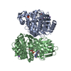 7s49C 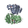 7s4cC  1wuuS S: Starting model for refinement C: citing same article ( |
|---|---|
| Similar structure data |


-Links
 PDBj
PDBj- Assembly
| Deposited unit | 
| ||||||||
|---|---|---|---|---|---|---|---|---|---|
| 1 | 
| ||||||||
| 2 | 
| ||||||||
| Unit cell |
|
-Components
-Protein / Sugars , 2 types, 8 molecules ABCD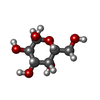
| #1: Protein | Mass: 42203.020 Da / Num. of mol.: 4 / Mutation: K252A, E253A Source method: isolated from a genetically manipulated source Source: (gene. exp.) Homo sapiens (human) / Gene: GALK1, GALK / Plasmid: pET21D / Production host:  #2: Sugar | ChemComp-GLA /  Type: D-saccharide, alpha linking / Mass: 180.156 Da / Num. of mol.: 4 / Source method: obtained synthetically / Formula: C6H12O6 Type: D-saccharide, alpha linking / Mass: 180.156 Da / Num. of mol.: 4 / Source method: obtained synthetically / Formula: C6H12O6 |
|---|
-Non-polymers , 5 types, 420 molecules 


| #3: Chemical | ChemComp-ADP /  Mass: 427.201 Da / Num. of mol.: 4 / Source method: obtained synthetically / Formula: C10H15N5O10P2 / Comment: ADP, energy-carrying molecule*YM Mass: 427.201 Da / Num. of mol.: 4 / Source method: obtained synthetically / Formula: C10H15N5O10P2 / Comment: ADP, energy-carrying molecule*YM#4: Chemical | ChemComp-PO4 /  Mass: 94.971 Da / Num. of mol.: 11 / Source method: obtained synthetically / Formula: PO4 Mass: 94.971 Da / Num. of mol.: 11 / Source method: obtained synthetically / Formula: PO4#5: Chemical | ChemComp-MG /  Mass: 24.305 Da / Num. of mol.: 4 / Source method: obtained synthetically / Formula: Mg Mass: 24.305 Da / Num. of mol.: 4 / Source method: obtained synthetically / Formula: Mg#6: Chemical | ChemComp-NA /  Mass: 22.990 Da / Num. of mol.: 4 / Source method: obtained synthetically / Formula: Na Mass: 22.990 Da / Num. of mol.: 4 / Source method: obtained synthetically / Formula: Na#7: Water | ChemComp-HOH / | Mass: 18.015 Da / Num. of mol.: 397 / Source method: isolated from a natural source / Formula: H2O |
|---|
-Details
| Has ligand of interest | N |
|---|---|
| Has protein modification | Y |
-Experimental details
-Experiment
| Experiment | Method: X-RAY DIFFRACTION / Number of used crystals: 1 |
|---|
- Sample preparation
| Crystal | Density Matthews: 3.39 Å3/Da / Density % sol: 63.67 % / Mosaicity: 0.518 ° / Mosaicity esd: 0.006 ° |
|---|---|
| Crystal grow | Temperature: 286 K / Method: vapor diffusion, sitting drop / pH: 6.8 Details: Seeding into drops containing GALK protein at 8 mg/ml, 2.3M Na/K pH6.8 |
-Data collection
| Diffraction | Mean temperature: 100 K / Serial crystal experiment: N | |||||||||||||||||||||||||||||||||||||||||||||||||||||||||||||||||||||||||||||||||||||||||||||||||||
|---|---|---|---|---|---|---|---|---|---|---|---|---|---|---|---|---|---|---|---|---|---|---|---|---|---|---|---|---|---|---|---|---|---|---|---|---|---|---|---|---|---|---|---|---|---|---|---|---|---|---|---|---|---|---|---|---|---|---|---|---|---|---|---|---|---|---|---|---|---|---|---|---|---|---|---|---|---|---|---|---|---|---|---|---|---|---|---|---|---|---|---|---|---|---|---|---|---|---|---|---|
| Diffraction source | Source: SYNCHROTRON / Site: SSRL / Beamline: BL7-1 / Wavelength: 1.1533 Å | |||||||||||||||||||||||||||||||||||||||||||||||||||||||||||||||||||||||||||||||||||||||||||||||||||
| Detector | Type: ADSC QUANTUM 1 / Detector: CCD / Date: Nov 21, 2013 | |||||||||||||||||||||||||||||||||||||||||||||||||||||||||||||||||||||||||||||||||||||||||||||||||||
| Radiation | Protocol: SINGLE WAVELENGTH / Monochromatic (M) / Laue (L): M / Scattering type: x-ray | |||||||||||||||||||||||||||||||||||||||||||||||||||||||||||||||||||||||||||||||||||||||||||||||||||
| Radiation wavelength | Wavelength: 1.1533 Å / Relative weight: 1 | |||||||||||||||||||||||||||||||||||||||||||||||||||||||||||||||||||||||||||||||||||||||||||||||||||
| Reflection | Resolution: 2.4→50 Å / Num. obs: 86404 / % possible obs: 95.7 % / Redundancy: 11.1 % / Rmerge(I) obs: 0.225 / Rpim(I) all: 0.069 / Rrim(I) all: 0.222 / Χ2: 1.216 / Net I/σ(I): 9 / Num. measured all: 955737 | |||||||||||||||||||||||||||||||||||||||||||||||||||||||||||||||||||||||||||||||||||||||||||||||||||
| Reflection shell | Diffraction-ID: 1
|
- Processing
| Software |
| |||||||||||||||||||||||||||||||||||||||||||||||||||||||||||||||||||||||||||||||||||||||||||||||||||||||||||||||||||||||||||||
|---|---|---|---|---|---|---|---|---|---|---|---|---|---|---|---|---|---|---|---|---|---|---|---|---|---|---|---|---|---|---|---|---|---|---|---|---|---|---|---|---|---|---|---|---|---|---|---|---|---|---|---|---|---|---|---|---|---|---|---|---|---|---|---|---|---|---|---|---|---|---|---|---|---|---|---|---|---|---|---|---|---|---|---|---|---|---|---|---|---|---|---|---|---|---|---|---|---|---|---|---|---|---|---|---|---|---|---|---|---|---|---|---|---|---|---|---|---|---|---|---|---|---|---|---|---|---|
| Refinement | Method to determine structure: MOLECULAR REPLACEMENT Starting model: 1wuu Resolution: 2.4→39.63 Å / Cor.coef. Fo:Fc: 0.964 / Cor.coef. Fo:Fc free: 0.94 / SU B: 16.248 / SU ML: 0.178 / SU R Cruickshank DPI: 0.2564 / Cross valid method: THROUGHOUT / σ(F): 0 / ESU R: 0.256 / ESU R Free: 0.216 / Stereochemistry target values: MAXIMUM LIKELIHOOD Details: HYDROGENS HAVE BEEN ADDED IN THE RIDING POSITIONS U VALUES : WITH TLS ADDED
| |||||||||||||||||||||||||||||||||||||||||||||||||||||||||||||||||||||||||||||||||||||||||||||||||||||||||||||||||||||||||||||
| Solvent computation | Ion probe radii: 0.8 Å / Shrinkage radii: 0.8 Å / VDW probe radii: 1.2 Å / Solvent model: MASK | |||||||||||||||||||||||||||||||||||||||||||||||||||||||||||||||||||||||||||||||||||||||||||||||||||||||||||||||||||||||||||||
| Displacement parameters | Biso max: 153.74 Å2 / Biso mean: 56.893 Å2 / Biso min: 30.76 Å2
| |||||||||||||||||||||||||||||||||||||||||||||||||||||||||||||||||||||||||||||||||||||||||||||||||||||||||||||||||||||||||||||
| Refinement step | Cycle: final / Resolution: 2.4→39.63 Å
| |||||||||||||||||||||||||||||||||||||||||||||||||||||||||||||||||||||||||||||||||||||||||||||||||||||||||||||||||||||||||||||
| Refine LS restraints |
| |||||||||||||||||||||||||||||||||||||||||||||||||||||||||||||||||||||||||||||||||||||||||||||||||||||||||||||||||||||||||||||
| LS refinement shell | Resolution: 2.4→2.462 Å / Rfactor Rfree error: 0 / Total num. of bins used: 20
| |||||||||||||||||||||||||||||||||||||||||||||||||||||||||||||||||||||||||||||||||||||||||||||||||||||||||||||||||||||||||||||
| Refinement TLS params. | Method: refined / Refine-ID: X-RAY DIFFRACTION
| |||||||||||||||||||||||||||||||||||||||||||||||||||||||||||||||||||||||||||||||||||||||||||||||||||||||||||||||||||||||||||||
| Refinement TLS group |
|
