[English] 日本語
 Yorodumi
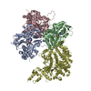
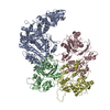
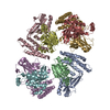
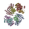
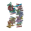
- PDB-7rb8: cryo-EM structure of the ADP state wild type myosin-15-F-actin complex -
+ Open data
- Basic information
| Entry | Database: PDB / ID: 7rb8 | ||||||||||||
|---|---|---|---|---|---|---|---|---|---|---|---|---|---|
| Title | cryo-EM structure of the ADP state wild type myosin-15-F-actin complex | ||||||||||||
 Components Components |
| ||||||||||||
 Keywords Keywords | MOTOR PROTEIN/ATP Binding Protein / myosin motor proteins / actin cytoskeleton / stereocilia / deafness / MOTOR PROTEIN / MOTOR PROTEIN-ATP Binding Protein complex | ||||||||||||
| Function / homology |  Function and homology information Function and homology informationStriated Muscle Contraction / stereocilium bundle / stereocilium / myosin complex / inner ear morphogenesis / cytoskeletal motor activity / striated muscle thin filament / response to light stimulus / skeletal muscle thin filament assembly / skeletal muscle fiber development ...Striated Muscle Contraction / stereocilium bundle / stereocilium / myosin complex / inner ear morphogenesis / cytoskeletal motor activity / striated muscle thin filament / response to light stimulus / skeletal muscle thin filament assembly / skeletal muscle fiber development / stress fiber / actin filament / locomotory behavior / sensory perception of sound / Hydrolases; Acting on acid anhydrides; Acting on acid anhydrides to facilitate cellular and subcellular movement / actin cytoskeleton / actin binding / hydrolase activity / ATP binding / cytosol Similarity search - Function | ||||||||||||
| Biological species |   | ||||||||||||
| Method | ELECTRON MICROSCOPY / helical reconstruction / cryo EM / Resolution: 3.63 Å | ||||||||||||
 Authors Authors | Gong, R. / Reynolds, M.J. / Gurel, P. / Alushin, G.M. | ||||||||||||
| Funding support |  United States, 3items United States, 3items
| ||||||||||||
 Citation Citation |  Journal: Sci Adv / Year: 2022 Journal: Sci Adv / Year: 2022Title: Structural basis for tunable control of actin dynamics by myosin-15 in mechanosensory stereocilia. Authors: Rui Gong / Fangfang Jiang / Zane G Moreland / Matthew J Reynolds / Santiago Espinosa de Los Reyes / Pinar Gurel / Arik Shams / James B Heidings / Michael R Bowl / Jonathan E Bird / Gregory M Alushin /   Abstract: The motor protein myosin-15 is necessary for the development and maintenance of mechanosensory stereocilia, and mutations in myosin-15 cause hereditary deafness. In addition to transporting actin ...The motor protein myosin-15 is necessary for the development and maintenance of mechanosensory stereocilia, and mutations in myosin-15 cause hereditary deafness. In addition to transporting actin regulatory machinery to stereocilia tips, myosin-15 directly nucleates actin filament ("F-actin") assembly, which is disrupted by a progressive hearing loss mutation (p.D1647G, ""). Here, we present cryo-electron microscopy structures of myosin-15 bound to F-actin, providing a framework for interpreting the impacts of deafness mutations on motor activity and actin nucleation. Rigor myosin-15 evokes conformational changes in F-actin yet maintains flexibility in actin's D-loop, which mediates inter-subunit contacts, while the mutant locks the D-loop in a single conformation. Adenosine diphosphate-bound myosin-15 also locks the D-loop, which correspondingly blunts actin-polymerization stimulation. We propose myosin-15 enhances polymerization by bridging actin protomers, regulating nucleation efficiency by modulating actin's structural plasticity in a myosin nucleotide state-dependent manner. This tunable regulation of actin polymerization could be harnessed to precisely control stereocilium height. #1:  Journal: Biorxiv / Year: 2021 Journal: Biorxiv / Year: 2021Title: Structural basis for tunable control of actin dynamics by myosin-15 in mechanosensory stereocilia Authors: Gong, R. / Jiang, F. / Moreland, Z.G. / Reynolds, M.J. / Gurel, P.S. / Shams, A. / Bowl, M.R. / Bird, J.E. / Alushin, G.M. | ||||||||||||
| History |
|
- Structure visualization
| Movie |
 Movie viewer Movie viewer |
|---|---|
| Structure viewer | Molecule:  Molmil Molmil Jmol/JSmol Jmol/JSmol |
- Downloads & links
- Download
| PDBx/mmCIF format |  7rb8.cif.gz 7rb8.cif.gz | 318 KB | Display |  PDBx/mmCIF format PDBx/mmCIF format |
|---|---|---|---|---|
| PDB format |  pdb7rb8.ent.gz pdb7rb8.ent.gz | 258.8 KB | Display |  PDB format PDB format |
| PDBx/mmJSON format |  7rb8.json.gz 7rb8.json.gz | Tree view |  PDBx/mmJSON format PDBx/mmJSON format | |
| Others |  Other downloads Other downloads |
-Validation report
| Summary document |  7rb8_validation.pdf.gz 7rb8_validation.pdf.gz | 1.3 MB | Display |  wwPDB validaton report wwPDB validaton report |
|---|---|---|---|---|
| Full document |  7rb8_full_validation.pdf.gz 7rb8_full_validation.pdf.gz | 1.3 MB | Display | |
| Data in XML |  7rb8_validation.xml.gz 7rb8_validation.xml.gz | 72.7 KB | Display | |
| Data in CIF |  7rb8_validation.cif.gz 7rb8_validation.cif.gz | 105.3 KB | Display | |
| Arichive directory |  https://data.pdbj.org/pub/pdb/validation_reports/rb/7rb8 https://data.pdbj.org/pub/pdb/validation_reports/rb/7rb8 ftp://data.pdbj.org/pub/pdb/validation_reports/rb/7rb8 ftp://data.pdbj.org/pub/pdb/validation_reports/rb/7rb8 | HTTPS FTP |
-Related structure data
| Related structure data |  24399MC  7r8vC  7r91C  7rb9C 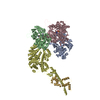 7udtC 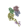 7uduC C: citing same article ( M: map data used to model this data |
|---|---|
| Similar structure data |
- Links
Links
- Assembly
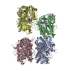
| Deposited unit | 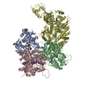
|
|---|---|
| 1 |
|
- Components
| #1: Protein | Mass: 41631.430 Da / Num. of mol.: 3 / Source method: isolated from a natural source / Source: (natural)  #2: Protein | | Mass: 78182.703 Da / Num. of mol.: 1 Source method: isolated from a genetically manipulated source Source: (gene. exp.)  Production host:  Spodoptera aff. frugiperda 2 RZ-2014 (butterflies/moths) Spodoptera aff. frugiperda 2 RZ-2014 (butterflies/moths)References: UniProt: Q9QZZ4 #3: Chemical | ChemComp-ADP / #4: Chemical | ChemComp-MG / Has ligand of interest | Y | |
|---|
-Experimental details
-Experiment
| Experiment | Method: ELECTRON MICROSCOPY |
|---|---|
| EM experiment | Aggregation state: FILAMENT / 3D reconstruction method: helical reconstruction |
- Sample preparation
| Component |
| ||||||||||||||||||||||||
|---|---|---|---|---|---|---|---|---|---|---|---|---|---|---|---|---|---|---|---|---|---|---|---|---|---|
| Molecular weight | Value: 5.4 kDa/nm / Experimental value: YES | ||||||||||||||||||||||||
| Source (natural) |
| ||||||||||||||||||||||||
| Buffer solution | pH: 7 Details: 10 mM imidazole pH 7.0, 50 mM KCl, 1 mM MgCl2, 1 mM EGTA, 0.5 mM DTT, 0.01% NaN3 | ||||||||||||||||||||||||
| Specimen | Embedding applied: NO / Shadowing applied: NO / Staining applied: NO / Vitrification applied: YES | ||||||||||||||||||||||||
| Specimen support | Grid material: GOLD / Grid mesh size: 300 divisions/in. / Grid type: C-flat-1.2/1.3 | ||||||||||||||||||||||||
| Vitrification | Instrument: LEICA EM GP / Cryogen name: ETHANE / Humidity: 100 % / Chamber temperature: 293 K |
- Electron microscopy imaging
| Experimental equipment |  Model: Titan Krios / Image courtesy: FEI Company |
|---|---|
| Microscopy | Model: FEI TITAN KRIOS |
| Electron gun | Electron source:  FIELD EMISSION GUN / Accelerating voltage: 300 kV / Illumination mode: FLOOD BEAM FIELD EMISSION GUN / Accelerating voltage: 300 kV / Illumination mode: FLOOD BEAM |
| Electron lens | Mode: BRIGHT FIELD / Nominal magnification: 29000 X / Nominal defocus max: 3500 nm / Nominal defocus min: 1500 nm / Alignment procedure: COMA FREE |
| Specimen holder | Cryogen: NITROGEN / Specimen holder model: FEI TITAN KRIOS AUTOGRID HOLDER |
| Image recording | Average exposure time: 10 sec. / Electron dose: 60 e/Å2 / Detector mode: SUPER-RESOLUTION / Film or detector model: GATAN K2 SUMMIT (4k x 4k) / Num. of grids imaged: 1 |
| Image scans | Movie frames/image: 40 / Used frames/image: 1-40 |
- Processing
| EM software |
| ||||||||||||||||||||||||||||||||||||||||
|---|---|---|---|---|---|---|---|---|---|---|---|---|---|---|---|---|---|---|---|---|---|---|---|---|---|---|---|---|---|---|---|---|---|---|---|---|---|---|---|---|---|
| CTF correction | Type: PHASE FLIPPING AND AMPLITUDE CORRECTION | ||||||||||||||||||||||||||||||||||||||||
| Helical symmerty | Angular rotation/subunit: -166.52 ° / Axial rise/subunit: 27.06 Å / Axial symmetry: C1 | ||||||||||||||||||||||||||||||||||||||||
| Particle selection | Num. of particles selected: 139547 | ||||||||||||||||||||||||||||||||||||||||
| 3D reconstruction | Resolution: 3.63 Å / Resolution method: FSC 0.143 CUT-OFF / Num. of particles: 125232 / Algorithm: BACK PROJECTION / Symmetry type: HELICAL | ||||||||||||||||||||||||||||||||||||||||
| Atomic model building | Protocol: RIGID BODY FIT / Space: REAL | ||||||||||||||||||||||||||||||||||||||||
| Atomic model building | PDB-ID: 6BNO |
 Movie
Movie Controller





 PDBj
PDBj


