 Movie
Movie Controller

[English] 日本語
 Yorodumi
- PDB-7rab: Crystal structure of a dodecameric multicopper oxidase from M. hy... -
+ Open data

- Basic information
| Entry | Database: PDB / ID: 7rab | ||||||
|---|---|---|---|---|---|---|---|
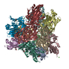
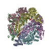
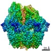
| Title | Crystal structure of a dodecameric multicopper oxidase from M. hydrothermalis in a cubic lattice | ||||||
 Components Components | multicopper oxidase | ||||||
 Keywords Keywords | OXIDOREDUCTASE / multicopper oxidase thermophile dodecamer laccase | ||||||
| Function / homology |  Function and homology information Function and homology informationnitrite reductase (NO-forming) / nitrite reductase (NO-forming) activity / copper ion binding Similarity search - Function | ||||||
| Biological species |  Marinithermus hydrothermalis (bacteria) Marinithermus hydrothermalis (bacteria) | ||||||
| Method |  X-RAY DIFFRACTION / SYNCHROTRON / MOLECULAR REPLACEMENT / molecular replacement / Resolution: 1.92 Å X-RAY DIFFRACTION / SYNCHROTRON / MOLECULAR REPLACEMENT / molecular replacement / Resolution: 1.92 Å | ||||||
 Authors Authors | Georgiadis, M.M. | ||||||
 Citation Citation | Journal: Acta Crystallogr D Struct Biol / Year: 2021 Title: Crystal structures of a dodecameric multicopper oxidase from Marinithermus hydrothermalis. Authors: Paavola, J.L. / Battistin, U. / Ogata, C.M. / Georgiadis, M.M. | ||||||
| History |
|
- Structure visualization
| Structure viewer | Molecule: MolmilJmol/JSmol |
|---|
- Downloads & links
-Download
| PDBx/mmCIF format | 7rab.cif.gz | 497 KB | Display | PDBx/mmCIF format |
|---|---|---|---|---|
| PDB format | pdb7rab.ent.gz | 403.9 KB | Display | PDB format |
| PDBx/mmJSON format | 7rab.json.gz | Tree view | PDBx/mmJSON format | |
| Others |  Other downloads Other downloads |
-Validation report
| Arichive directory | https://data.pdbj.org/pub/pdb/validation_reports/ra/7rabftp://data.pdbj.org/pub/pdb/validation_reports/ra/7rab | HTTPS FTP |
|---|
-Related structure data


-Links
 PDBj
PDBj

- Assembly
| Deposited unit | 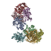
| ||||||||
|---|---|---|---|---|---|---|---|---|---|
| 1 | 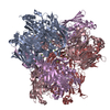
| ||||||||
| 2 | 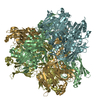
| ||||||||
| Unit cell |
| ||||||||
| Components on special symmetry positions |
|
-Components
| #1: Protein | Mass: 39424.773 Da / Num. of mol.: 8 Source method: isolated from a genetically manipulated source Source: (gene. exp.) Marinithermus hydrothermalis (bacteria)Gene: Marky_0543 / Production host: #2: Chemical | ChemComp-CU /   Mass: 63.546 Da / Num. of mol.: 16 / Source method: obtained synthetically / Formula: Cu / Feature type: SUBJECT OF INVESTIGATION Mass: 63.546 Da / Num. of mol.: 16 / Source method: obtained synthetically / Formula: Cu / Feature type: SUBJECT OF INVESTIGATION#3: Chemical | ChemComp-MG /   Mass: 24.305 Da / Num. of mol.: 8 / Source method: obtained synthetically / Formula: Mg Mass: 24.305 Da / Num. of mol.: 8 / Source method: obtained synthetically / Formula: Mg#4: Water | ChemComp-HOH / |  Mass: 18.015 Da / Num. of mol.: 619 / Source method: isolated from a natural source / Formula: H2O Mass: 18.015 Da / Num. of mol.: 619 / Source method: isolated from a natural source / Formula: H2OHas ligand of interest | Y | |
|---|
-Experimental details
-Experiment
| Experiment | Method: X-RAY DIFFRACTION / Number of used crystals: 1 |
|---|
- Sample preparation
| Crystal | Density Matthews: 2.98 Å3/Da / Density % sol: 58.71 % |
|---|---|
| Crystal grow | Temperature: 293 K / Method: vapor diffusion, hanging drop Details: 0.2 M ammonium sulfate, 0.1 M HEPES 7.5, 25% PEG 335 PH range: 7.5 |
-Data collection
| Diffraction | Mean temperature: 100 K / Serial crystal experiment: N | |||||||||||||||||||||||||||
|---|---|---|---|---|---|---|---|---|---|---|---|---|---|---|---|---|---|---|---|---|---|---|---|---|---|---|---|---|
| Diffraction source | Source: SYNCHROTRON / Site: APS  / Beamline: 31-ID / Wavelength: 1.37623 Å / Beamline: 31-ID / Wavelength: 1.37623 Å | |||||||||||||||||||||||||||
| Detector | Type: DECTRIS PILATUS3 6M / Detector: PIXEL / Date: Jul 19, 2019 | |||||||||||||||||||||||||||
| Radiation | Protocol: SINGLE WAVELENGTH / Monochromatic (M) / Laue (L): M / Scattering type: x-ray | |||||||||||||||||||||||||||
| Radiation wavelength | Wavelength: 1.37623 Å / Relative weight: 1 | |||||||||||||||||||||||||||
| Reflection | Resolution: 1.915→129.47 Å / Num. obs: 284806 / % possible obs: 99.7 % / Redundancy: 39 % / Biso Wilson estimate: 27.16 Å2 / CC1/2: 1 / Rmerge(I) obs: 0.17 / Rpim(I) all: 0.028 / Rrim(I) all: 0.173 / Net I/σ(I): 10.6 | |||||||||||||||||||||||||||
| Reflection shell | Diffraction-ID: 1 / % possible all: 99.8
|
-Phasing
| Phasing | Method: molecular replacement |
|---|
- Processing
| Software |
| ||||||||||||||||||||||||||||||||||||||||||||||||||||||||||||||||||||||||||||||||||||||||||||||||||||||||||||||||||||||||||||||||||||||||||||||||||||||||||||||||||||||||||||||||||||||||||
|---|---|---|---|---|---|---|---|---|---|---|---|---|---|---|---|---|---|---|---|---|---|---|---|---|---|---|---|---|---|---|---|---|---|---|---|---|---|---|---|---|---|---|---|---|---|---|---|---|---|---|---|---|---|---|---|---|---|---|---|---|---|---|---|---|---|---|---|---|---|---|---|---|---|---|---|---|---|---|---|---|---|---|---|---|---|---|---|---|---|---|---|---|---|---|---|---|---|---|---|---|---|---|---|---|---|---|---|---|---|---|---|---|---|---|---|---|---|---|---|---|---|---|---|---|---|---|---|---|---|---|---|---|---|---|---|---|---|---|---|---|---|---|---|---|---|---|---|---|---|---|---|---|---|---|---|---|---|---|---|---|---|---|---|---|---|---|---|---|---|---|---|---|---|---|---|---|---|---|---|---|---|---|---|---|---|---|---|
| Refinement | Method to determine structure: MOLECULAR REPLACEMENT Starting model: 3GDC_A Resolution: 1.92→56.06 Å / SU ML: 0.22 / Cross valid method: THROUGHOUT / σ(F): 1.33 / Phase error: 43.45 / Stereochemistry target values: ML
| ||||||||||||||||||||||||||||||||||||||||||||||||||||||||||||||||||||||||||||||||||||||||||||||||||||||||||||||||||||||||||||||||||||||||||||||||||||||||||||||||||||||||||||||||||||||||||
| Solvent computation | Shrinkage radii: 0.9 Å / VDW probe radii: 1.11 Å / Solvent model: FLAT BULK SOLVENT MODEL | ||||||||||||||||||||||||||||||||||||||||||||||||||||||||||||||||||||||||||||||||||||||||||||||||||||||||||||||||||||||||||||||||||||||||||||||||||||||||||||||||||||||||||||||||||||||||||
| Displacement parameters | Biso max: 84.94 Å2 / Biso mean: 30.3317 Å2 / Biso min: 15.65 Å2 | ||||||||||||||||||||||||||||||||||||||||||||||||||||||||||||||||||||||||||||||||||||||||||||||||||||||||||||||||||||||||||||||||||||||||||||||||||||||||||||||||||||||||||||||||||||||||||
| Refinement step | Cycle: final / Resolution: 1.92→56.06 Å
| ||||||||||||||||||||||||||||||||||||||||||||||||||||||||||||||||||||||||||||||||||||||||||||||||||||||||||||||||||||||||||||||||||||||||||||||||||||||||||||||||||||||||||||||||||||||||||
| Refine LS restraints |
| ||||||||||||||||||||||||||||||||||||||||||||||||||||||||||||||||||||||||||||||||||||||||||||||||||||||||||||||||||||||||||||||||||||||||||||||||||||||||||||||||||||||||||||||||||||||||||
| LS refinement shell | Refine-ID: X-RAY DIFFRACTION / Rfactor Rfree error: 0
|
