 Movie
Movie Controller

[English] 日本語
 Yorodumi
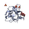
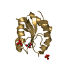
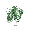
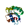
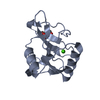
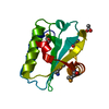
- PDB-7r8x: Crystal Structure of the LNK SH2 Domain in Complex with an EPOR p... -
+ Open data

- Basic information
| Entry | Database: PDB / ID: 7r8x | ||||||
|---|---|---|---|---|---|---|---|
| Title | Crystal Structure of the LNK SH2 Domain in Complex with an EPOR pY454 Phosphopeptide | ||||||
 Components Components |
| ||||||
 Keywords Keywords | SIGNALING PROTEIN / LNK / SH2B3 / JAK/STAT / MPNs | ||||||
| Function / homology |  Function and homology information Function and homology informationnegative regulation of Kit signaling pathway / monocyte homeostasis / stem cell factor receptor binding / Regulation of KIT signaling / Negative regulation of FLT3 / negative regulation of chemokine-mediated signaling pathway / Factors involved in megakaryocyte development and platelet production / thrombopoietin-mediated signaling pathway / negative regulation of receptor signaling pathway via JAK-STAT / negative regulation of platelet aggregation ...negative regulation of Kit signaling pathway / monocyte homeostasis / stem cell factor receptor binding / Regulation of KIT signaling / Negative regulation of FLT3 / negative regulation of chemokine-mediated signaling pathway / Factors involved in megakaryocyte development and platelet production / thrombopoietin-mediated signaling pathway / negative regulation of receptor signaling pathway via JAK-STAT / negative regulation of platelet aggregation / neutrophil homeostasis / cellular response to chemokine / cellular response to interleukin-3 / negative regulation of receptor signaling pathway via STAT / embryonic hemopoiesis / megakaryocyte development / erythrocyte development / hemopoiesis / hematopoietic stem cell differentiation / negative regulation of MAPK cascade / negative regulation of phosphatidylinositol 3-kinase/protein kinase B signal transduction / protein tyrosine kinase binding / signaling receptor complex adaptor activity / intracellular signal transduction / negative regulation of cell population proliferation Similarity search - Function | ||||||
| Biological species |   Homo sapiens (human) Homo sapiens (human) | ||||||
| Method |  X-RAY DIFFRACTION / SYNCHROTRON / MOLECULAR REPLACEMENT / Resolution: 2.3 Å X-RAY DIFFRACTION / SYNCHROTRON / MOLECULAR REPLACEMENT / Resolution: 2.3 Å | ||||||
 Authors Authors | Morris, R. / Kershaw, N.J. / Babon, J.J. | ||||||
| Funding support |  Australia, 1items Australia, 1items
| ||||||
 Citation Citation | Journal: Nat Commun / Year: 2021 Title: Structural and functional analysis of target recognition by the lymphocyte adaptor protein LNK. Authors: Morris, R. / Zhang, Y. / Ellyard, J.I. / Vinuesa, C.G. / Murphy, J.M. / Laktyushin, A. / Kershaw, N.J. / Babon, J.J. | ||||||
| History |
|
- Structure visualization
| Structure viewer | Molecule: MolmilJmol/JSmol |
|---|
- Downloads & links
-Download
| PDBx/mmCIF format | 7r8x.cif.gz | 39.3 KB | Display | PDBx/mmCIF format |
|---|---|---|---|---|
| PDB format | pdb7r8x.ent.gz | 24.2 KB | Display | PDB format |
| PDBx/mmJSON format | 7r8x.json.gz | Tree view | PDBx/mmJSON format | |
| Others |  Other downloads Other downloads |
-Validation report
| Summary document | 7r8x_validation.pdf.gz | 423.9 KB | Display | wwPDB validaton report |
|---|---|---|---|---|
| Full document | 7r8x_full_validation.pdf.gz | 424 KB | Display | |
| Data in XML | 7r8x_validation.xml.gz | 6.8 KB | Display | |
| Data in CIF | 7r8x_validation.cif.gz | 8.1 KB | Display | |
| Arichive directory | https://data.pdbj.org/pub/pdb/validation_reports/r8/7r8xftp://data.pdbj.org/pub/pdb/validation_reports/r8/7r8x | HTTPS FTP |
-Related structure data
| Related structure data | 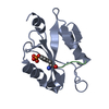 7r8wC 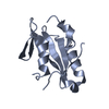 2hdvS C: citing same article ( S: Starting model for refinement |
|---|---|
| Similar structure data |


-Links
 PDBj
PDBj


- Assembly
| Deposited unit | 
| ||||||||||
|---|---|---|---|---|---|---|---|---|---|---|---|
| 1 |
| ||||||||||
| Unit cell |
|
-Components
| #1: Protein | Mass: 12682.654 Da / Num. of mol.: 1 / Fragment: UNP residues 328-438 Source method: isolated from a genetically manipulated source Source: (gene. exp.)  |
|---|---|
| #2: Protein/peptide | Mass: 1065.177 Da / Num. of mol.: 1 / Source method: obtained synthetically / Source: (synth.) Homo sapiens (human) |
| #3: Water | ChemComp-HOH /  Mass: 18.015 Da / Num. of mol.: 13 / Source method: isolated from a natural source / Formula: H2O Mass: 18.015 Da / Num. of mol.: 13 / Source method: isolated from a natural source / Formula: H2O |
| Has ligand of interest | Y |
| Has protein modification | Y |
-Experimental details
-Experiment
| Experiment | Method: X-RAY DIFFRACTION / Number of used crystals: 1 |
|---|
- Sample preparation
| Crystal | Density Matthews: 2.97 Å3/Da / Density % sol: 58.57 % |
|---|---|
| Crystal grow | Temperature: 277.15 K / Method: vapor diffusion, hanging drop / pH: 9.5 Details: 20% PEG8000, 0.05 M magnesium acetate, 0.1 M Tris, ph 8.5 |
-Data collection
| Diffraction | Mean temperature: 100 K / Serial crystal experiment: N |
|---|---|
| Diffraction source | Source: SYNCHROTRON / Site: Australian Synchrotron / Beamline: MX2 / Wavelength: 0.95374 Å |
| Detector | Type: DECTRIS EIGER X 16M / Detector: PIXEL / Date: Jun 21, 2018 |
| Radiation | Protocol: SINGLE WAVELENGTH / Monochromatic (M) / Laue (L): M / Scattering type: x-ray |
| Radiation wavelength | Wavelength: 0.95374 Å / Relative weight: 1 |
| Reflection | Resolution: 2.3→33.9 Å / Num. obs: 7318 / % possible obs: 99.78 % / Redundancy: 9.6 % / Biso Wilson estimate: 53.5 Å2 / CC1/2: 1 / CC star: 1 / Rmerge(I) obs: 0.04408 / Rpim(I) all: 0.01481 / Rrim(I) all: 0.04655 / Net I/σ(I): 31 |
| Reflection shell | Resolution: 2.301→2.383 Å / Rmerge(I) obs: 0.4924 / Mean I/σ(I) obs: 2.81 / Num. unique obs: 724 / CC1/2: 0.904 / CC star: 0.974 / Rrim(I) all: 0.539 / % possible all: 99.45 |
- Processing
| Software |
| ||||||||||||||||||||||||||||||||||||||||||
|---|---|---|---|---|---|---|---|---|---|---|---|---|---|---|---|---|---|---|---|---|---|---|---|---|---|---|---|---|---|---|---|---|---|---|---|---|---|---|---|---|---|---|---|
| Refinement | Method to determine structure: MOLECULAR REPLACEMENT Starting model: PDB entry 2HDV Resolution: 2.3→33.9 Å / SU ML: 0.34 / Cross valid method: THROUGHOUT / σ(F): 1.36 / Phase error: 29.29 / Stereochemistry target values: ML
| ||||||||||||||||||||||||||||||||||||||||||
| Solvent computation | Shrinkage radii: 0.9 Å / VDW probe radii: 1.11 Å / Solvent model: FLAT BULK SOLVENT MODEL | ||||||||||||||||||||||||||||||||||||||||||
| Displacement parameters | Biso max: 93.72 Å2 / Biso mean: 53.8189 Å2 / Biso min: 30.34 Å2 | ||||||||||||||||||||||||||||||||||||||||||
| Refinement step | Cycle: final / Resolution: 2.3→33.9 Å
| ||||||||||||||||||||||||||||||||||||||||||
| LS refinement shell | Refine-ID: X-RAY DIFFRACTION / Rfactor Rfree error: 0 / Total num. of bins used: 5
|
