 Movie
Movie Controller

[English] 日本語
 Yorodumi
- PDB-1fwv: CRYSTAL STRUCTURE OF THE CYSTEINE-RICH DOMAIN OF MANNOSE RECEPTOR... -
+ Open data

- Basic information
| Entry | Database: PDB / ID: 1fwv | |||||||||
|---|---|---|---|---|---|---|---|---|---|---|
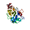
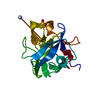
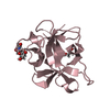
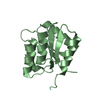
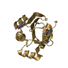
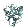
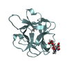
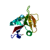
| Title | CRYSTAL STRUCTURE OF THE CYSTEINE-RICH DOMAIN OF MANNOSE RECEPTOR COMPLEXED WITH 3-SO4-LEWIS(A) | |||||||||
 Components Components | CYSTEINE-RICH DOMAIN OF MANNOSE RECEPTOR | |||||||||
 Keywords Keywords | SUGAR BINDING PROTEIN / beta trefoil / mannose receptor / sulfated carbohydrate | |||||||||
| Function / homology |  Function and homology information Function and homology informationCross-presentation of soluble exogenous antigens (endosomes) / cargo receptor activity / D-mannose binding / cellular response to interleukin-4 / receptor-mediated endocytosis / cellular response to type II interferon / transmembrane signaling receptor activity / cellular response to lipopolysaccharide / signaling receptor activity / endosome membrane ...Cross-presentation of soluble exogenous antigens (endosomes) / cargo receptor activity / D-mannose binding / cellular response to interleukin-4 / receptor-mediated endocytosis / cellular response to type II interferon / transmembrane signaling receptor activity / cellular response to lipopolysaccharide / signaling receptor activity / endosome membrane / cell surface / plasma membrane Similarity search - Function | |||||||||
| Biological species |  | |||||||||
| Method |  X-RAY DIFFRACTION / MOLECULAR REPLACEMENT / Resolution: 2.2 Å X-RAY DIFFRACTION / MOLECULAR REPLACEMENT / Resolution: 2.2 Å | |||||||||
 Authors Authors | Liu, Y. / Misulovin, Z. / Bjorkman, P.J. | |||||||||
 Citation Citation | Journal: J.Mol.Biol. / Year: 2001 Title: The molecular mechanism of sulfated carbohydrate recognition by the cysteine-rich domain of mannose receptor. Authors: Liu, Y. / Misulovin, Z. / Bjorkman, P.J. | |||||||||
| History |
|
- Structure visualization
| Structure viewer | Molecule: MolmilJmol/JSmol |
|---|
- Downloads & links
-Download
| PDBx/mmCIF format | 1fwv.cif.gz | 45.4 KB | Display | PDBx/mmCIF format |
|---|---|---|---|---|
| PDB format | pdb1fwv.ent.gz | 30.2 KB | Display | PDB format |
| PDBx/mmJSON format | 1fwv.json.gz | Tree view | PDBx/mmJSON format | |
| Others |  Other downloads Other downloads |
-Validation report
| Arichive directory | https://data.pdbj.org/pub/pdb/validation_reports/fw/1fwvftp://data.pdbj.org/pub/pdb/validation_reports/fw/1fwv | HTTPS FTP |
|---|
-Related structure data


-Links
 PDBj
PDBj


- Assembly
| Deposited unit | 
| ||||||||||
|---|---|---|---|---|---|---|---|---|---|---|---|
| 1 |
| ||||||||||
| Unit cell |
|
-Components
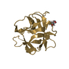
| #1: Protein | Mass: 15445.328 Da / Num. of mol.: 1 / Fragment: N-TERMINAL DOMAIN OF MANNOSE RECEPTOR Source method: isolated from a genetically manipulated source Details: COMPLEXED WITH 3-SO4-LEWIS(A) / Source: (gene. exp.) |
|---|---|
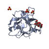
| #2: Polysaccharide | 3-O-sulfo-beta-D-galactopyranose-(1-3)-[alpha-L-fucopyranose-(1-4)]methyl 2-acetamido-2-deoxy-beta- ...3-O-sulfo-beta-D-galactopyranose-(1-3)-[alpha-L-fucopyranose-(1-4)]methyl 2-acetamido-2-deoxy-beta-D-glucopyranoside Source method: isolated from a genetically manipulated source |
| #3: Water | ChemComp-HOH /  Mass: 18.015 Da / Num. of mol.: 127 / Source method: isolated from a natural source / Formula: H2O Mass: 18.015 Da / Num. of mol.: 127 / Source method: isolated from a natural source / Formula: H2O |
| Has protein modification | Y |
-Experimental details
-Experiment
| Experiment | Method: X-RAY DIFFRACTION / Number of used crystals: 1 |
|---|
- Sample preparation
| Crystal | Density Matthews: 2.63 Å3/Da / Density % sol: 53.21 % | ||||||||||||||||||||||||||||||||||||
|---|---|---|---|---|---|---|---|---|---|---|---|---|---|---|---|---|---|---|---|---|---|---|---|---|---|---|---|---|---|---|---|---|---|---|---|---|---|
| Crystal grow | Temperature: 277 K / Method: vapor diffusion, hanging drop / pH: 7 Details: PEG 8000, (NH4)2SO4, pH 6.5-7.5 , pH 7.0, VAPOR DIFFUSION, HANGING DROP, temperature 277K | ||||||||||||||||||||||||||||||||||||
| Crystal grow | *PLUS Temperature: 4 ℃ / pH: 7.4 | ||||||||||||||||||||||||||||||||||||
| Components of the solutions | *PLUS
|
-Data collection
| Diffraction | Mean temperature: 120 K |
|---|---|
| Diffraction source | Source: ROTATING ANODE / Type: RIGAKU RU200 / Wavelength: 1.5418 |
| Detector | Type: RIGAKU RAXIS II / Detector: IMAGE PLATE / Date: Feb 21, 2000 |
| Radiation | Protocol: SINGLE WAVELENGTH / Monochromatic (M) / Laue (L): M / Scattering type: x-ray |
| Radiation wavelength | Wavelength: 1.5418 Å / Relative weight: 1 |
| Reflection | Resolution: 2.2→25 Å / Num. all: 8862 / Num. obs: 8538 / % possible obs: 96.5 % / Observed criterion σ(F): 0 / Observed criterion σ(I): 0 / Redundancy: 7.6 % / Biso Wilson estimate: 32.7 Å2 / Rmerge(I) obs: 0.034 / Net I/σ(I): 31 |
| Reflection shell | Resolution: 2.2→2.28 Å / Redundancy: 2.9 % / Rmerge(I) obs: 0.081 / % possible all: 89.1 |
| Reflection | *PLUS Num. measured all: 64935 |
| Reflection shell | *PLUS % possible obs: 89.1 % / Rmerge(I) obs: 0.085 / Mean I/σ(I) obs: 12.4 |
- Processing
| Software |
| |||||||||||||||||||||||||
|---|---|---|---|---|---|---|---|---|---|---|---|---|---|---|---|---|---|---|---|---|---|---|---|---|---|---|
| Refinement | Method to determine structure: MOLECULAR REPLACEMENT / Resolution: 2.2→25 Å / Cross valid method: THROUGHOUT / σ(F): 0 / σ(I): 0 / Stereochemistry target values: Engh & Huber
| |||||||||||||||||||||||||
| Refinement step | Cycle: LAST / Resolution: 2.2→25 Å
| |||||||||||||||||||||||||
| Refine LS restraints |
| |||||||||||||||||||||||||
| Software | *PLUS Name: CNS / Classification: refinement | |||||||||||||||||||||||||
| Refinement | *PLUS Highest resolution: 2.2 Å / Lowest resolution: 25 Å / σ(F): 0 / % reflection Rfree: 6.3 % / Rfactor obs: 0.208 / Rfactor Rfree: 0.23 | |||||||||||||||||||||||||
| Solvent computation | *PLUS | |||||||||||||||||||||||||
| Displacement parameters | *PLUS |
