 ムービー
ムービー コントローラー


+ データを開く

- 基本情報
基本情報
| 登録情報 | データベース: PDB / ID: 7r66 | ||||||
|---|---|---|---|---|---|---|---|
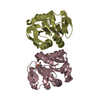
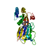
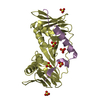
| タイトル | Structure of Pfp1 protease from Thermococcus thioreducens: large unit cell at 1.44 A resolution | ||||||
 要素 要素 | Peptidase | ||||||
 キーワード キーワード | HYDROLASE / intracellular / disulfide bonds / catalytic triad / proteasome / bacteria / supersite | ||||||
| 機能・相同性 | : / Deglycase PfpI / PfpI endopeptidase domain profile. / DJ-1/PfpI / DJ-1/PfpI family / Class I glutamine amidotransferase-like / PHOSPHATE ION / Peptidase 機能・相同性情報 機能・相同性情報 | ||||||
| 生物種 |   Thermococcus thioreducens (古細菌) Thermococcus thioreducens (古細菌) | ||||||
| 手法 |  X線回折 / シンクロトロン / 分子置換 / 解像度: 1.44 Å X線回折 / シンクロトロン / 分子置換 / 解像度: 1.44 Å | ||||||
 データ登録者 データ登録者 | McPherson, A. | ||||||
 引用 引用 | ジャーナル: Acta Crystallogr. / 年: 2017 タイトル: Structure of the Pfp1 protease from a hypothermophilic archer, Thermococcus thioreducens, in two crystal forms 著者: Larson, S.B. / McPherson, A. | ||||||
| 履歴 |
|
- 構造の表示
構造の表示
| 構造ビューア | 分子: MolmilJmol/JSmol |
|---|
- ダウンロードとリンク
-ダウンロード
| PDBx/mmCIF形式 | 7r66.cif.gz | 503.3 KB | 表示 | PDBx/mmCIF形式 |
|---|---|---|---|---|
| PDB形式 | pdb7r66.ent.gz | 351.6 KB | 表示 | PDB形式 |
| PDBx/mmJSON形式 | 7r66.json.gz | ツリー表示 | PDBx/mmJSON形式 | |
| その他 |  その他のダウンロード その他のダウンロード |
-検証レポート
| 文書・要旨 | 7r66_validation.pdf.gz | 477.9 KB | 表示 | wwPDB検証レポート |
|---|---|---|---|---|
| 文書・詳細版 | 7r66_full_validation.pdf.gz | 483.9 KB | 表示 | |
| XML形式データ | 7r66_validation.xml.gz | 33.9 KB | 表示 | |
| CIF形式データ | 7r66_validation.cif.gz | 48.8 KB | 表示 | |
| アーカイブディレクトリ | https://data.pdbj.org/pub/pdb/validation_reports/r6/7r66ftp://data.pdbj.org/pub/pdb/validation_reports/r6/7r66 | HTTPS FTP |
-関連構造データ
| 関連構造データ |  5txwS S: 精密化の開始モデル |
|---|---|
| 類似構造データ |


-リンク
 PDBj
PDBj


- 集合体
集合体
| 登録構造単位 | 
| ||||||||||||
|---|---|---|---|---|---|---|---|---|---|---|---|---|---|
| 1 | 
| ||||||||||||
| 2 | 
| ||||||||||||
| 単位格子 |
|
-要素
| #1: タンパク質 | 分子量: 18461.092 Da / 分子数: 4 / 由来タイプ: 組換発現 由来: (組換発現) Thermococcus thioreducens (古細菌)遺伝子: AMR53_10535 / 発現宿主:  #2: 化合物 | ChemComp-MPD / (   分子量: 118.174 Da / 分子数: 4 / 由来タイプ: 合成 / 式: C6H14O2 / コメント: 沈殿剤*YM 分子量: 118.174 Da / 分子数: 4 / 由来タイプ: 合成 / 式: C6H14O2 / コメント: 沈殿剤*YM#3: 化合物 |   分子量: 94.971 Da / 分子数: 2 / 由来タイプ: 合成 / 式: PO4 分子量: 94.971 Da / 分子数: 2 / 由来タイプ: 合成 / 式: PO4#4: 水 | ChemComp-HOH / |  分子量: 18.015 Da / 分子数: 598 / 由来タイプ: 天然 / 式: H2O 分子量: 18.015 Da / 分子数: 598 / 由来タイプ: 天然 / 式: H2O研究の焦点であるリガンドがあるか | N | |
|---|
-実験情報
-実験
| 実験 | 手法: X線回折 / 使用した結晶の数: 1 |
|---|
- 試料調製
試料調製
| 結晶 | マシュー密度: 2.48 Å3/Da / 溶媒含有率: 50 % / 解説: hexagonal prisms |
|---|---|
| 結晶化 | 温度: 298 K / 手法: 蒸気拡散法, シッティングドロップ法 / pH: 6.5 詳細: Sitting drop crystallization using Cryschem plates. Drops were equal amounts of a 25 mg / ml protein stock solution in water and the reservoir solution. The reservoir was 1.4 M sodium citrate ...詳細: Sitting drop crystallization using Cryschem plates. Drops were equal amounts of a 25 mg / ml protein stock solution in water and the reservoir solution. The reservoir was 1.4 M sodium citrate at pH 6.5. Crystallization was at room temperature. Drops were 10 ul total starting volume, reservoirs 0.6 ml. PH範囲: 6.0 - 7.0 |
-データ収集
| 回折 | 平均測定温度: 173 K / Serial crystal experiment: N |
|---|---|
| 放射光源 | 由来: シンクロトロン / サイト: ALS  / ビームライン: 8.3.1 / 波長: 1.1158 Å / ビームライン: 8.3.1 / 波長: 1.1158 Å |
| 検出器 | タイプ: DECTRIS EIGER X 500K / 検出器: PIXEL / 日付: 2019年6月15日 |
| 放射 | プロトコル: SINGLE WAVELENGTH / 単色(M)・ラウエ(L): M / 散乱光タイプ: x-ray |
| 放射波長 | 波長: 1.1158 Å / 相対比: 1 |
| 反射 | 解像度: 1.38→75 Å / Num. obs: 141230 / % possible obs: 100 % / 冗長度: 47 % / Biso Wilson estimate: 19.95 Å2 / CC1/2: 0.999 / Rmerge(I) obs: 0.216 / Rpim(I) all: 0.44 / Rrim(I) all: 0.221 / Rsym value: 0.21 / Net I/σ(I): 11.9 |
| 反射 シェル | 解像度: 1.38→1.4 Å / 冗長度: 19.8 % / Mean I/σ(I) obs: 0.3 / Num. unique obs: 7080 / CC1/2: 0.073 / % possible all: 100 |
- 解析
解析
| ソフトウェア |
| |||||||||||||||||||||||||||||||||||||||||||||||||||||||||||||||||||||||||||||||||||||||||||||||||||||||||||||||||||||||||||||||||||||||||||||||||||||||||||||||||||||||||||||||||||||||||||||||||||||||||||||||||||||||||
|---|---|---|---|---|---|---|---|---|---|---|---|---|---|---|---|---|---|---|---|---|---|---|---|---|---|---|---|---|---|---|---|---|---|---|---|---|---|---|---|---|---|---|---|---|---|---|---|---|---|---|---|---|---|---|---|---|---|---|---|---|---|---|---|---|---|---|---|---|---|---|---|---|---|---|---|---|---|---|---|---|---|---|---|---|---|---|---|---|---|---|---|---|---|---|---|---|---|---|---|---|---|---|---|---|---|---|---|---|---|---|---|---|---|---|---|---|---|---|---|---|---|---|---|---|---|---|---|---|---|---|---|---|---|---|---|---|---|---|---|---|---|---|---|---|---|---|---|---|---|---|---|---|---|---|---|---|---|---|---|---|---|---|---|---|---|---|---|---|---|---|---|---|---|---|---|---|---|---|---|---|---|---|---|---|---|---|---|---|---|---|---|---|---|---|---|---|---|---|---|---|---|---|---|---|---|---|---|---|---|---|---|---|---|---|---|---|---|---|
| 精密化 | 構造決定の手法: 分子置換 開始モデル: 5TXW 解像度: 1.44→68.69 Å / SU ML: 0.1423 / 交差検証法: FREE R-VALUE / σ(F): 1.96 / 位相誤差: 18.5948 立体化学のターゲット値: GeoStd + Monomer Library + CDL v1.2
| |||||||||||||||||||||||||||||||||||||||||||||||||||||||||||||||||||||||||||||||||||||||||||||||||||||||||||||||||||||||||||||||||||||||||||||||||||||||||||||||||||||||||||||||||||||||||||||||||||||||||||||||||||||||||
| 溶媒の処理 | 減衰半径: 0.9 Å / VDWプローブ半径: 1.11 Å / 溶媒モデル: FLAT BULK SOLVENT MODEL | |||||||||||||||||||||||||||||||||||||||||||||||||||||||||||||||||||||||||||||||||||||||||||||||||||||||||||||||||||||||||||||||||||||||||||||||||||||||||||||||||||||||||||||||||||||||||||||||||||||||||||||||||||||||||
| 原子変位パラメータ | Biso mean: 27.88 Å2 | |||||||||||||||||||||||||||||||||||||||||||||||||||||||||||||||||||||||||||||||||||||||||||||||||||||||||||||||||||||||||||||||||||||||||||||||||||||||||||||||||||||||||||||||||||||||||||||||||||||||||||||||||||||||||
| 精密化ステップ | サイクル: LAST / 解像度: 1.44→68.69 Å
| |||||||||||||||||||||||||||||||||||||||||||||||||||||||||||||||||||||||||||||||||||||||||||||||||||||||||||||||||||||||||||||||||||||||||||||||||||||||||||||||||||||||||||||||||||||||||||||||||||||||||||||||||||||||||
| 拘束条件 |
| |||||||||||||||||||||||||||||||||||||||||||||||||||||||||||||||||||||||||||||||||||||||||||||||||||||||||||||||||||||||||||||||||||||||||||||||||||||||||||||||||||||||||||||||||||||||||||||||||||||||||||||||||||||||||
| LS精密化 シェル |
|
