 Movie
Movie Controller

[English] 日本語
 Yorodumi
- PDB-5txw: Structure of Pfp1 protease from Thermococcus thioreducens: large ... -
+ Open data

- Basic information
| Entry | Database: PDB / ID: 5txw | ||||||
|---|---|---|---|---|---|---|---|
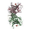
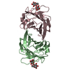
| Title | Structure of Pfp1 protease from Thermococcus thioreducens: large cell H3 crystal form | ||||||
 Components Components | Peptidase | ||||||
 Keywords Keywords | HYDROLASE / protease / hexamer / thermophile / noncrystallographic symmetry | ||||||
| Function / homology |  Function and homology information Function and homology information: / Deglycase PfpI / PfpI endopeptidase domain profile. / DJ-1/PfpI / DJ-1/PfpI family / Class I glutamine amidotransferase (GATase) domain / Class I glutamine amidotransferase-like / Rossmann fold / 3-Layer(aba) Sandwich / Alpha Beta Similarity search - Domain/homology | ||||||
| Biological species |   Thermococcus thioreducens (archaea) Thermococcus thioreducens (archaea) | ||||||
| Method |  X-RAY DIFFRACTION / MOLECULAR REPLACEMENT / Resolution: 1.86 Å X-RAY DIFFRACTION / MOLECULAR REPLACEMENT / Resolution: 1.86 Å | ||||||
 Authors Authors | Larson, S.B. / McPherson, A. | ||||||
 Citation Citation | Journal: Acta Crystallogr D Struct Biol / Year: 2017 Title: The structure of the Pfp1 protease from the hyperthermophilic archaeon Thermococcus thioreducens in two crystal forms. Authors: Larson, S.B. / McPherson, A. | ||||||
| History |
|
- Structure visualization
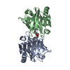
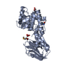
| Structure viewer | Molecule: MolmilJmol/JSmol |
|---|
- Downloads & links
-Download
| PDBx/mmCIF format | 5txw.cif.gz | 278.9 KB | Display | PDBx/mmCIF format |
|---|---|---|---|---|
| PDB format | pdb5txw.ent.gz | 230.8 KB | Display | PDB format |
| PDBx/mmJSON format | 5txw.json.gz | Tree view | PDBx/mmJSON format | |
| Others |  Other downloads Other downloads |
-Validation report
| Arichive directory | https://data.pdbj.org/pub/pdb/validation_reports/tx/5txwftp://data.pdbj.org/pub/pdb/validation_reports/tx/5txw | HTTPS FTP |
|---|
-Related structure data
| Related structure data |  5tw0C  1g2iS C: citing same article ( S: Starting model for refinement |
|---|---|
| Similar structure data |


-Links
 PDBj
PDBj

- Assembly
| Deposited unit | 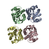
| ||||||||
|---|---|---|---|---|---|---|---|---|---|
| 1 | 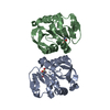
| ||||||||
| 2 | 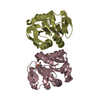
| ||||||||
| Unit cell |
|
-Components
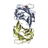
| #1: Protein | Mass: 18461.092 Da / Num. of mol.: 4 Source method: isolated from a genetically manipulated source Source: (gene. exp.) Thermococcus thioreducens (archaea) / Gene: AMR53_10535 / Production host:  #2: Water | ChemComp-HOH / |  Mass: 18.015 Da / Num. of mol.: 639 / Source method: isolated from a natural source / Formula: H2O Mass: 18.015 Da / Num. of mol.: 639 / Source method: isolated from a natural source / Formula: H2O |
|---|
-Experimental details
-Experiment
| Experiment | Method: X-RAY DIFFRACTION / Number of used crystals: 5 |
|---|
- Sample preparation
| Crystal | Density Matthews: 2.528 Å3/Da / Density % sol: 51.3 % / Description: large hexagonal prisms |
|---|---|
| Crystal grow | Temperature: 293 K / Method: vapor diffusion, sitting drop / pH: 6.5 Details: 1.4 M Sodium Citrate, pH 6.5, vapor diffusion, sitting drop Temp details: Room temperature |
-Data collection
| Diffraction | Mean temperature: 298 K / Ambient temp details: Room temperature |
|---|---|
| Diffraction source | Source: ROTATING ANODE / Type: RIGAKU MICROMAX-007 HF / Wavelength: 1.54187 Å |
| Detector | Type: RIGAKU SATURN 944+ / Detector: CCD / Date: Apr 10, 2013 / Details: Osmic mirrors |
| Radiation | Protocol: SINGLE WAVELENGTH / Monochromatic (M) / Laue (L): M / Scattering type: x-ray |
| Radiation wavelength | Wavelength: 1.54187 Å / Relative weight: 1 |
| Reflection | Resolution: 1.86→38.22 Å / Num. obs: 52126 / % possible obs: 86.3 % / Observed criterion σ(F): 0 / Observed criterion σ(I): 0 / Redundancy: 8.8 % / Biso Wilson estimate: 15.88 Å2 / CC1/2: 0.992 / Rmerge(I) obs: 0.123 / Net I/σ(I): 11.9 |
| Reflection shell | Resolution: 1.86→1.9 Å / Redundancy: 1 % / Rmerge(I) obs: 0.583 / Mean I/σ(I) obs: 0.6 / CC1/2: 0.485 / % possible all: 33.5 |
- Processing
| Software |
| ||||||||||||||||||||||||||||||||||||||||||||||||||||||||||||||||||||||||||||||||||||||||||||||||||||||||||||||||||||||||||||||||||||||||||||||||||||||||||||||||||||||||||||||||||||||
|---|---|---|---|---|---|---|---|---|---|---|---|---|---|---|---|---|---|---|---|---|---|---|---|---|---|---|---|---|---|---|---|---|---|---|---|---|---|---|---|---|---|---|---|---|---|---|---|---|---|---|---|---|---|---|---|---|---|---|---|---|---|---|---|---|---|---|---|---|---|---|---|---|---|---|---|---|---|---|---|---|---|---|---|---|---|---|---|---|---|---|---|---|---|---|---|---|---|---|---|---|---|---|---|---|---|---|---|---|---|---|---|---|---|---|---|---|---|---|---|---|---|---|---|---|---|---|---|---|---|---|---|---|---|---|---|---|---|---|---|---|---|---|---|---|---|---|---|---|---|---|---|---|---|---|---|---|---|---|---|---|---|---|---|---|---|---|---|---|---|---|---|---|---|---|---|---|---|---|---|---|---|---|---|
| Refinement | Method to determine structure: MOLECULAR REPLACEMENT Starting model: 1G2I Resolution: 1.86→38.22 Å / Cor.coef. Fo:Fc: 0.975 / Cor.coef. Fo:Fc free: 0.957 / SU B: 3.597 / SU ML: 0.096 / SU R Cruickshank DPI: 0.1236 / Cross valid method: THROUGHOUT / σ(F): 0 / ESU R: 0.124 / ESU R Free: 0.12 / SU Rfree Cruickshank DPI: 0.1203 / Details: HYDROGENS HAVE BEEN USED IF PRESENT IN THE INPUT
| ||||||||||||||||||||||||||||||||||||||||||||||||||||||||||||||||||||||||||||||||||||||||||||||||||||||||||||||||||||||||||||||||||||||||||||||||||||||||||||||||||||||||||||||||||||||
| Solvent computation | Ion probe radii: 0.8 Å / Shrinkage radii: 0.8 Å / VDW probe radii: 1.2 Å / Solvent model: Babinet bulk solvent modeling | ||||||||||||||||||||||||||||||||||||||||||||||||||||||||||||||||||||||||||||||||||||||||||||||||||||||||||||||||||||||||||||||||||||||||||||||||||||||||||||||||||||||||||||||||||||||
| Displacement parameters | Biso mean: 20.02 Å2
| ||||||||||||||||||||||||||||||||||||||||||||||||||||||||||||||||||||||||||||||||||||||||||||||||||||||||||||||||||||||||||||||||||||||||||||||||||||||||||||||||||||||||||||||||||||||
| Refinement step | Cycle: 1 / Resolution: 1.86→38.22 Å
| ||||||||||||||||||||||||||||||||||||||||||||||||||||||||||||||||||||||||||||||||||||||||||||||||||||||||||||||||||||||||||||||||||||||||||||||||||||||||||||||||||||||||||||||||||||||
| Refine LS restraints |
|