 Movie
Movie Controller


+ Open data

- Basic information
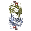
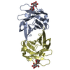
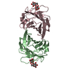
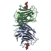
| Entry | Database: PDB / ID: 1ww7 | ||||||
|---|---|---|---|---|---|---|---|
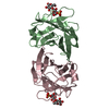
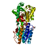
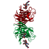
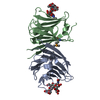
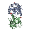
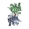
| Title | Agrocybe cylindracea galectin (Ligand-free) | ||||||
 Components Components | galectin | ||||||
 Keywords Keywords | SUGAR BINDING PROTEIN / Agrocybe cylindracea galectin / fungal galectin / carbohydrate recognition domain / X-ray crystallographic analysis / sulfate ion | ||||||
| Function / homology |  Function and homology information Function and homology informationDNA nuclease activity / Hydrolases; Acting on ester bonds; Endodeoxyribonucleases producing 5'-phosphomonoesters / polysaccharide binding / positive regulation of apoptotic process / hydrolase activity / apoptotic process Similarity search - Function | ||||||
| Biological species |  Agrocybe cylindracea (poplar fieldcap) Agrocybe cylindracea (poplar fieldcap) | ||||||
| Method |  X-RAY DIFFRACTION / MOLECULAR REPLACEMENT / Resolution: 1.9 Å X-RAY DIFFRACTION / MOLECULAR REPLACEMENT / Resolution: 1.9 Å | ||||||
 Authors Authors | Ban, M. / Yoon, H.J. / Demirkan, E. / Utsumi, S. / Mikami, B. / Yagi, F. | ||||||
 Citation Citation | Journal: J.Mol.Biol. / Year: 2005 Title: Structural Basis of a Fungal Galectin from Agrocybe cylindracea for Recognizing Sialoconjugate Authors: Ban, M. / Yoon, H.J. / Demirkan, E. / Utsumi, S. / Mikami, B. / Yagi, F. | ||||||
| History |
|
- Structure visualization
| Structure viewer | Molecule: MolmilJmol/JSmol |
|---|
- Downloads & links
-Download
| PDBx/mmCIF format | 1ww7.cif.gz | 138.9 KB | Display | PDBx/mmCIF format |
|---|---|---|---|---|
| PDB format | pdb1ww7.ent.gz | 109.7 KB | Display | PDB format |
| PDBx/mmJSON format | 1ww7.json.gz | Tree view | PDBx/mmJSON format | |
| Others |  Other downloads Other downloads |
-Validation report
| Arichive directory | https://data.pdbj.org/pub/pdb/validation_reports/ww/1ww7ftp://data.pdbj.org/pub/pdb/validation_reports/ww/1ww7 | HTTPS FTP |
|---|
-Related structure data


-Links
 PDBj
PDBj- Assembly
| Deposited unit | 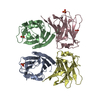
| ||||||||
|---|---|---|---|---|---|---|---|---|---|
| 1 | 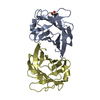
| ||||||||
| 2 | 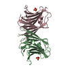
| ||||||||
| Unit cell |
|
-Components
| #1: Protein | Mass: 16908.744 Da / Num. of mol.: 4 / Source method: isolated from a natural source / Source: (natural) Agrocybe cylindracea (poplar fieldcap) / References: UniProt: Q6WY08*PLUS#2: Chemical | ChemComp-SO4 /   Mass: 96.063 Da / Num. of mol.: 4 / Source method: obtained synthetically / Formula: SO4 Mass: 96.063 Da / Num. of mol.: 4 / Source method: obtained synthetically / Formula: SO4#3: Water | ChemComp-HOH / |  Mass: 18.015 Da / Num. of mol.: 453 / Source method: isolated from a natural source / Formula: H2O Mass: 18.015 Da / Num. of mol.: 453 / Source method: isolated from a natural source / Formula: H2O |
|---|
-Experimental details
-Experiment
| Experiment | Method: X-RAY DIFFRACTION / Number of used crystals: 1 |
|---|
- Sample preparation
| Crystal | Density Matthews: 2.4 Å3/Da / Density % sol: 47.5 % |
|---|---|
| Crystal grow | Temperature: 293 K / Method: vapor diffusion, hanging drop / pH: 7.1 Details: ammomium sulfate, pipes, pH 7.1, VAPOR DIFFUSION, HANGING DROP, temperature 293K |
-Data collection
| Diffraction | Mean temperature: 293 K |
|---|---|
| Diffraction source | Source: ROTATING ANODE / Type: MACSCIENCE / Wavelength: 1.5418 Å |
| Detector | Type: SIEMENS HI-STAR / Detector: AREA DETECTOR / Date: Feb 10, 1999 / Details: carbon monochromater |
| Radiation | Monochromator: carbon monochromater / Protocol: SINGLE WAVELENGTH / Monochromatic (M) / Laue (L): M / Scattering type: x-ray |
| Radiation wavelength | Wavelength: 1.5418 Å / Relative weight: 1 |
| Reflection | Resolution: 1.9→41.7 Å / Num. all: 53735 / Num. obs: 51284 / % possible obs: 95.4 % / Observed criterion σ(F): 0 / Observed criterion σ(I): 1 / Redundancy: 5.32 % / Biso Wilson estimate: 1.3 Å2 / Rsym value: 0.109 / Net I/σ(I): 11.09 |
| Reflection shell | Resolution: 1.9→1.97 Å / Redundancy: 2.95 % / Mean I/σ(I) obs: 1.86 / Num. unique all: 4562 / Rsym value: 0.464 / % possible all: 85.9 |
- Processing
| Software |
| ||||||||||||||||||||||||||||||||||||
|---|---|---|---|---|---|---|---|---|---|---|---|---|---|---|---|---|---|---|---|---|---|---|---|---|---|---|---|---|---|---|---|---|---|---|---|---|---|
| Refinement | Method to determine structure: MOLECULAR REPLACEMENT / Resolution: 1.9→10 Å / Rfactor Rfree error: 0.004 / Data cutoff high absF: 503729.2 / Data cutoff low absF: 0 / Isotropic thermal model: RESTRAINED / Cross valid method: THROUGHOUT / σ(F): 0 / Stereochemistry target values: Engh & Huber
| ||||||||||||||||||||||||||||||||||||
| Solvent computation | Solvent model: FLAT MODEL / Bsol: 77.9027 Å2 / ksol: 0.367265 e/Å3 | ||||||||||||||||||||||||||||||||||||
| Displacement parameters | Biso mean: 18 Å2
| ||||||||||||||||||||||||||||||||||||
| Refine analyze |
| ||||||||||||||||||||||||||||||||||||
| Refinement step | Cycle: LAST / Resolution: 1.9→10 Å
| ||||||||||||||||||||||||||||||||||||
| Refine LS restraints |
| ||||||||||||||||||||||||||||||||||||
| LS refinement shell | Resolution: 1.9→2.02 Å / Rfactor Rfree error: 0.014 / Total num. of bins used: 6
| ||||||||||||||||||||||||||||||||||||
| Xplor file |
|
