 Movie
Movie Controller

[English] 日本語
 Yorodumi
- PDB-3m3o: Crystal Structure of Agrocybe aegerita lectin AAL mutant R85A com... -
+ Open data

- Basic information
| Entry | Database: PDB / ID: 3m3o | |||||||||
|---|---|---|---|---|---|---|---|---|---|---|
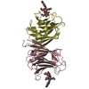
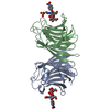
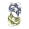
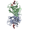
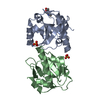
| Title | Crystal Structure of Agrocybe aegerita lectin AAL mutant R85A complexed with p-Nitrophenyl TF disaccharide | |||||||||
 Components Components | Anti-tumor lectin | |||||||||
 Keywords Keywords | HYDROLASE / galectin / AAL / muitant / Thomsen-Friedenreich antigen / Apoptosis / Lectin / Nuclease / GAL-BETA-1 / 3-GALNAC-ALPHA-O-P-Nitrophenyl | |||||||||
| Function / homology |  Function and homology information Function and homology informationDNA nuclease activity / Hydrolases; Acting on ester bonds; Endodeoxyribonucleases producing 5'-phosphomonoesters / polysaccharide binding / positive regulation of apoptotic process / hydrolase activity / apoptotic process Similarity search - Function | |||||||||
| Biological species |  Agrocybe aegerita (fungus) Agrocybe aegerita (fungus) | |||||||||
| Method |  X-RAY DIFFRACTION / MOLECULAR REPLACEMENT / Resolution: 2.1 Å X-RAY DIFFRACTION / MOLECULAR REPLACEMENT / Resolution: 2.1 Å | |||||||||
 Authors Authors | Feng, L. / Li, D. / Wang, D. | |||||||||
 Citation Citation | Journal: Faseb J. / Year: 2010 Title: Structural insights into the recognition mechanism between an antitumor galectin AAL and the Thomsen-Friedenreich antigen Authors: Feng, L. / Sun, H. / Zhang, Y. / Li, D.F. / Wang, D.C. | |||||||||
| History |
|
- Structure visualization
| Structure viewer | Molecule: MolmilJmol/JSmol |
|---|
- Downloads & links
-Download
| PDBx/mmCIF format | 3m3o.cif.gz | 48.4 KB | Display | PDBx/mmCIF format |
|---|---|---|---|---|
| PDB format | pdb3m3o.ent.gz | 31.8 KB | Display | PDB format |
| PDBx/mmJSON format | 3m3o.json.gz | Tree view | PDBx/mmJSON format | |
| Others |  Other downloads Other downloads |
-Validation report
| Arichive directory | https://data.pdbj.org/pub/pdb/validation_reports/m3/3m3oftp://data.pdbj.org/pub/pdb/validation_reports/m3/3m3o | HTTPS FTP |
|---|
-Related structure data
| Related structure data | 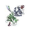 3afkC  3m3cC 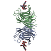 3m3eC 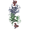 3m3qC 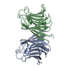 2zglS C: citing same article ( S: Starting model for refinement |
|---|---|
| Similar structure data |

-Links
 PDBj
PDBj
- Assembly
| Deposited unit | 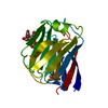
| ||||||||
|---|---|---|---|---|---|---|---|---|---|
| 1 | 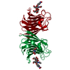
| ||||||||
| Unit cell |
|
-Components
| #1: Protein | Mass: 16891.684 Da / Num. of mol.: 1 / Mutation: R85A Source method: isolated from a genetically manipulated source Source: (gene. exp.) Agrocybe aegerita (fungus) / Gene: AAL / Plasmid: pET22b / Production host:  References: UniProt: Q6WY08, Hydrolases; Acting on ester bonds; Endodeoxyribonucleases producing 5'-phosphomonoesters |
|---|---|
| #2: Polysaccharide | beta-D-galactopyranose-(1-3)-2-acetamido-2-deoxy-alpha-D-galactopyranose / Thomsen-Friedenreich antigen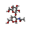  Source method: isolated from a genetically manipulated source Details: oligosaccharide / References: Thomsen-Friedenreich antigen |
| #3: Chemical | ChemComp-NPO / 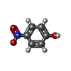  Mass: 139.109 Da / Num. of mol.: 1 / Source method: obtained synthetically / Formula: C6H5NO3 Mass: 139.109 Da / Num. of mol.: 1 / Source method: obtained synthetically / Formula: C6H5NO3 |
| #4: Chemical | ChemComp-BTB /   Mass: 209.240 Da / Num. of mol.: 1 / Source method: obtained synthetically / Formula: C8H19NO5 / Comment: pH buffer*YM Mass: 209.240 Da / Num. of mol.: 1 / Source method: obtained synthetically / Formula: C8H19NO5 / Comment: pH buffer*YM |
| #5: Water | ChemComp-HOH /  Mass: 18.015 Da / Num. of mol.: 55 / Source method: isolated from a natural source / Formula: H2O Mass: 18.015 Da / Num. of mol.: 55 / Source method: isolated from a natural source / Formula: H2O |
| Sequence details | IT IS AN ALLELE GENE OF THE GENE IN THE GENBANK DATABASE. |
-Experimental details
-Experiment
| Experiment | Method: X-RAY DIFFRACTION / Number of used crystals: 1 |
|---|
- Sample preparation
| Crystal | Density Matthews: 1.94 Å3/Da / Density % sol: 36.58 % |
|---|---|
| Crystal grow | Temperature: 298 K / Method: hanging drop / pH: 5.5 Details: 25% PEG3350, 0.2M LiCl, 5% acetone, pH 5.5, hanging drop, temperature 298K |
-Data collection
| Diffraction | Mean temperature: 100 K |
|---|---|
| Diffraction source | Source: ROTATING ANODE / Type: RIGAKU FR-E+ SUPERBRIGHT / Wavelength: 1.5418 Å |
| Detector | Type: RIGAKU RAXIS IV / Detector: IMAGE PLATE / Date: Aug 25, 2009 |
| Radiation | Protocol: SINGLE WAVELENGTH / Monochromatic (M) / Laue (L): M / Scattering type: x-ray |
| Radiation wavelength | Wavelength: 1.5418 Å / Relative weight: 1 |
| Reflection | Resolution: 2.1→36.893 Å / Num. all: 8217 / Num. obs: 8216 / % possible obs: 100 % / Redundancy: 5.9 % / Biso Wilson estimate: 22.1 Å2 / Rmerge(I) obs: 0.047 / Rsym value: 0.047 / Net I/σ(I): 10.4 |
| Reflection shell | Resolution: 2.1→2.21 Å / Redundancy: 5.9 % / Rmerge(I) obs: 0.279 / Mean I/σ(I) obs: 5.7 / Num. unique all: 1183 / Rsym value: 0.279 / % possible all: 100 |
- Processing
| Software |
| ||||||||||||||||||||||||||||||||||||||||||||||||||||||
|---|---|---|---|---|---|---|---|---|---|---|---|---|---|---|---|---|---|---|---|---|---|---|---|---|---|---|---|---|---|---|---|---|---|---|---|---|---|---|---|---|---|---|---|---|---|---|---|---|---|---|---|---|---|---|---|
| Refinement | Method to determine structure: MOLECULAR REPLACEMENT Starting model: 2ZGL Resolution: 2.1→36.86 Å / FOM work R set: 0.803 / Isotropic thermal model: Isotropic / Cross valid method: THROUGHOUT / σ(F): 0 / Stereochemistry target values: Engh & Huber
| ||||||||||||||||||||||||||||||||||||||||||||||||||||||
| Solvent computation | Bsol: 47.237 Å2 | ||||||||||||||||||||||||||||||||||||||||||||||||||||||
| Displacement parameters | Biso mean: 37.242 Å2
| ||||||||||||||||||||||||||||||||||||||||||||||||||||||
| Refine analyze |
| ||||||||||||||||||||||||||||||||||||||||||||||||||||||
| Refinement step | Cycle: LAST / Resolution: 2.1→36.86 Å
| ||||||||||||||||||||||||||||||||||||||||||||||||||||||
| Refine LS restraints |
| ||||||||||||||||||||||||||||||||||||||||||||||||||||||
| LS refinement shell | Refine-ID: X-RAY DIFFRACTION / Total num. of bins used: 8
| ||||||||||||||||||||||||||||||||||||||||||||||||||||||
| Xplor file |
|
