 Movie
Movie Controller

[English] 日本語
 Yorodumi
- PDB-7r1e: Mosquitocidal Cry11Ba determined at pH 10.4 from naturally-occurr... -
+ Open data

- Basic information
| Entry | Database: PDB / ID: 7r1e | |||||||||||||||||||||
|---|---|---|---|---|---|---|---|---|---|---|---|---|---|---|---|---|---|---|---|---|---|---|
| Title | Mosquitocidal Cry11Ba determined at pH 10.4 from naturally-occurring nanocrystals by Serial femtosecond crystallography | |||||||||||||||||||||
 Components Components | Pesticidal crystal protein Cry11Ba | |||||||||||||||||||||
 Keywords Keywords | TOXIN / naturally-occurring crystals mosquitocidal toxin serial femtosecond crystallography | |||||||||||||||||||||
| Function / homology |  Function and homology information Function and homology informationsymbiont-mediated killing of host cell / sporulation resulting in formation of a cellular spore / toxin activity Similarity search - Function | |||||||||||||||||||||
| Biological species |  | |||||||||||||||||||||
| Method |  X-RAY DIFFRACTION / FREE ELECTRON LASER / FOURIER SYNTHESIS / Resolution: 2.65 Å X-RAY DIFFRACTION / FREE ELECTRON LASER / FOURIER SYNTHESIS / Resolution: 2.65 Å | |||||||||||||||||||||
 Authors Authors | Colletier, J.-P. / Sawaya, M.R. / Schibrowsky, N.A. / Cascio, D. / Rodriguez, J.A. | |||||||||||||||||||||
| Funding support |  France, 6items France, 6items
| |||||||||||||||||||||
 Citation Citation | Journal: Nat Commun / Year: 2022 Title: De novo determination of mosquitocidal Cry11Aa and Cry11Ba structures from naturally-occurring nanocrystals. Authors: Tetreau, G. / Sawaya, M.R. / De Zitter, E. / Andreeva, E.A. / Banneville, A.S. / Schibrowsky, N.A. / Coquelle, N. / Brewster, A.S. / Grunbein, M.L. / Kovacs, G.N. / Hunter, M.S. / Kloos, M. ...Authors: Tetreau, G. / Sawaya, M.R. / De Zitter, E. / Andreeva, E.A. / Banneville, A.S. / Schibrowsky, N.A. / Coquelle, N. / Brewster, A.S. / Grunbein, M.L. / Kovacs, G.N. / Hunter, M.S. / Kloos, M. / Sierra, R.G. / Schiro, G. / Qiao, P. / Stricker, M. / Bideshi, D. / Young, I.D. / Zala, N. / Engilberge, S. / Gorel, A. / Signor, L. / Teulon, J.M. / Hilpert, M. / Foucar, L. / Bielecki, J. / Bean, R. / de Wijn, R. / Sato, T. / Kirkwood, H. / Letrun, R. / Batyuk, A. / Snigireva, I. / Fenel, D. / Schubert, R. / Canfield, E.J. / Alba, M.M. / Laporte, F. / Despres, L. / Bacia, M. / Roux, A. / Chapelle, C. / Riobe, F. / Maury, O. / Ling, W.L. / Boutet, S. / Mancuso, A. / Gutsche, I. / Girard, E. / Barends, T.R.M. / Pellequer, J.L. / Park, H.W. / Laganowsky, A.D. / Rodriguez, J. / Burghammer, M. / Shoeman, R.L. / Doak, R.B. / Weik, M. / Sauter, N.K. / Federici, B. / Cascio, D. / Schlichting, I. / Colletier, J.P. | |||||||||||||||||||||
| History |
|
- Structure visualization
| Structure viewer | Molecule: MolmilJmol/JSmol |
|---|
- Downloads & links
-Download
| PDBx/mmCIF format | 7r1e.cif.gz | 556.5 KB | Display | PDBx/mmCIF format |
|---|---|---|---|---|
| PDB format | pdb7r1e.ent.gz | 408.4 KB | Display | PDB format |
| PDBx/mmJSON format | 7r1e.json.gz | Tree view | PDBx/mmJSON format | |
| Others |  Other downloads Other downloads |
-Validation report
| Summary document | 7r1e_validation.pdf.gz | 454.5 KB | Display | wwPDB validaton report |
|---|---|---|---|---|
| Full document | 7r1e_full_validation.pdf.gz | 463.2 KB | Display | |
| Data in XML | 7r1e_validation.xml.gz | 42.7 KB | Display | |
| Data in CIF | 7r1e_validation.cif.gz | 58.6 KB | Display | |
| Arichive directory | https://data.pdbj.org/pub/pdb/validation_reports/r1/7r1eftp://data.pdbj.org/pub/pdb/validation_reports/r1/7r1e | HTTPS FTP |
-Related structure data
| Related structure data | 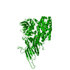 7qx4C 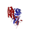 7qx5C 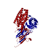 7qx6C  7qx7C  7qydSC S: Starting model for refinement C: citing same article ( |
|---|---|
| Similar structure data | |
| Experimental dataset #1 | Data reference: 10.11577/1873154 / Data set type: diffraction image data |
-Links
 PDBj
PDBj- Assembly
| Deposited unit | 
| ||||||||||||
|---|---|---|---|---|---|---|---|---|---|---|---|---|---|
| 1 |
| ||||||||||||
| Unit cell |
|
-Components
| #1: Protein | Mass: 81412.625 Da / Num. of mol.: 2 Source method: isolated from a genetically manipulated source Source: (gene. exp.) Gene: cry11Ba, cry11B, cryXIB(a) Production host: References: UniProt: Q45730 #2: Chemical |   Mass: 92.094 Da / Num. of mol.: 2 / Source method: obtained synthetically / Formula: C3H8O3 Mass: 92.094 Da / Num. of mol.: 2 / Source method: obtained synthetically / Formula: C3H8O3#3: Water | ChemComp-HOH / |  Mass: 18.015 Da / Num. of mol.: 119 / Source method: isolated from a natural source / Formula: H2O Mass: 18.015 Da / Num. of mol.: 119 / Source method: isolated from a natural source / Formula: H2OHas ligand of interest | N | Has protein modification | Y | |
|---|
-Experimental details
-Experiment
| Experiment | Method: X-RAY DIFFRACTION / Number of used crystals: 1 |
|---|
- Sample preparation
| Crystal | Density Matthews: 2.33 Å3/Da / Density % sol: 47.29 % |
|---|---|
| Crystal grow | Temperature: 303 K / Method: in cell / pH: 10.4 / Details: crystallized in vivo |
-Data collection
| Diffraction | Mean temperature: 298 K / Serial crystal experiment: Y |
|---|---|
| Diffraction source | Source: FREE ELECTRON LASER / Site: SLAC LCLS  / Beamline: CXI / Wavelength: 1.303351 Å / Beamline: CXI / Wavelength: 1.303351 Å |
| Detector | Type: CS-PAD CXI-2 / Detector: PIXEL / Date: Nov 15, 2018 |
| Radiation | Protocol: SINGLE WAVELENGTH / Monochromatic (M) / Laue (L): M / Scattering type: x-ray |
| Radiation wavelength | Wavelength: 1.303351 Å / Relative weight: 1 |
| Reflection | Resolution: 2.65→35.72 Å / Num. obs: 45243 / % possible obs: 99.4 % / Redundancy: 77 % / Biso Wilson estimate: 32.2 Å2 / CC1/2: 0.985 / R split: 0.224 / Net I/σ(I): 3.98 |
| Reflection shell | Resolution: 2.65→2.7 Å / Redundancy: 21 % / Mean I/σ(I) obs: 1.02 / Num. unique obs: 2204 / CC1/2: 0.152 / R split: 0.84 / % possible all: 100 |
| Serial crystallography measurement | Focal spot size: 1 µm2 / Pulse duration: 41 fsec. / Pulse energy: 1.1 µJ / Pulse photon energy: 9.5 keV / Source distance: 140 m / Source size: 1 µm2 / XFEL pulse repetition rate: 120 Hz |
| Serial crystallography sample delivery | Description: microfluidic electrokinetic sample holder / Method: injection |
| Serial crystallography sample delivery injection | Carrier solvent: 50% glycerol, 0.1M CAPS buffer pH 10.4 / Description: microfluidic electrokinetic sample holder / Flow rate: 1 µL/min |
| Serial crystallography data reduction | Lattices indexed: 15689 |
- Processing
| Software |
| |||||||||||||||||||||||||||||||||||||||||||||||||||||||||||||||||||||||||||||||||||||||||||||||||||||||||||||||||||||||||||||||||||||||||||||||||||||||||||||||||||||||||||||||||||||||||||||||||||||||||||||||||||||||||
|---|---|---|---|---|---|---|---|---|---|---|---|---|---|---|---|---|---|---|---|---|---|---|---|---|---|---|---|---|---|---|---|---|---|---|---|---|---|---|---|---|---|---|---|---|---|---|---|---|---|---|---|---|---|---|---|---|---|---|---|---|---|---|---|---|---|---|---|---|---|---|---|---|---|---|---|---|---|---|---|---|---|---|---|---|---|---|---|---|---|---|---|---|---|---|---|---|---|---|---|---|---|---|---|---|---|---|---|---|---|---|---|---|---|---|---|---|---|---|---|---|---|---|---|---|---|---|---|---|---|---|---|---|---|---|---|---|---|---|---|---|---|---|---|---|---|---|---|---|---|---|---|---|---|---|---|---|---|---|---|---|---|---|---|---|---|---|---|---|---|---|---|---|---|---|---|---|---|---|---|---|---|---|---|---|---|---|---|---|---|---|---|---|---|---|---|---|---|---|---|---|---|---|---|---|---|---|---|---|---|---|---|---|---|---|---|---|---|---|
| Refinement | Method to determine structure: FOURIER SYNTHESIS Starting model: 7QYD Resolution: 2.65→35.72 Å / SU ML: 0.4167 / Cross valid method: FREE R-VALUE / σ(F): 1.34 / Phase error: 26.2186 Stereochemistry target values: GeoStd + Monomer Library + CDL v1.2
| |||||||||||||||||||||||||||||||||||||||||||||||||||||||||||||||||||||||||||||||||||||||||||||||||||||||||||||||||||||||||||||||||||||||||||||||||||||||||||||||||||||||||||||||||||||||||||||||||||||||||||||||||||||||||
| Solvent computation | Shrinkage radii: 0.9 Å / VDW probe radii: 1.1 Å / Solvent model: FLAT BULK SOLVENT MODEL | |||||||||||||||||||||||||||||||||||||||||||||||||||||||||||||||||||||||||||||||||||||||||||||||||||||||||||||||||||||||||||||||||||||||||||||||||||||||||||||||||||||||||||||||||||||||||||||||||||||||||||||||||||||||||
| Displacement parameters | Biso mean: 45.39 Å2 | |||||||||||||||||||||||||||||||||||||||||||||||||||||||||||||||||||||||||||||||||||||||||||||||||||||||||||||||||||||||||||||||||||||||||||||||||||||||||||||||||||||||||||||||||||||||||||||||||||||||||||||||||||||||||
| Refinement step | Cycle: LAST / Resolution: 2.65→35.72 Å
| |||||||||||||||||||||||||||||||||||||||||||||||||||||||||||||||||||||||||||||||||||||||||||||||||||||||||||||||||||||||||||||||||||||||||||||||||||||||||||||||||||||||||||||||||||||||||||||||||||||||||||||||||||||||||
| Refine LS restraints |
| |||||||||||||||||||||||||||||||||||||||||||||||||||||||||||||||||||||||||||||||||||||||||||||||||||||||||||||||||||||||||||||||||||||||||||||||||||||||||||||||||||||||||||||||||||||||||||||||||||||||||||||||||||||||||
| LS refinement shell |
| |||||||||||||||||||||||||||||||||||||||||||||||||||||||||||||||||||||||||||||||||||||||||||||||||||||||||||||||||||||||||||||||||||||||||||||||||||||||||||||||||||||||||||||||||||||||||||||||||||||||||||||||||||||||||
| Refinement TLS params. | Method: refined / Refine-ID: X-RAY DIFFRACTION
| |||||||||||||||||||||||||||||||||||||||||||||||||||||||||||||||||||||||||||||||||||||||||||||||||||||||||||||||||||||||||||||||||||||||||||||||||||||||||||||||||||||||||||||||||||||||||||||||||||||||||||||||||||||||||
| Refinement TLS group | Refine-ID: X-RAY DIFFRACTION
|
