 Movie
Movie Controller

[English] 日本語
 Yorodumi
- PDB-7qjt: Crystal structure of a cutinase enzyme from Thermobifida cellulos... -
+ Open data

- Basic information
| Entry | Database: PDB / ID: 7qjt | ||||||
|---|---|---|---|---|---|---|---|
| Title | Crystal structure of a cutinase enzyme from Thermobifida cellulosilytica TB100 (711) | ||||||
 Components Components | cutinase (711) | ||||||
 Keywords Keywords | HYDROLASE / plastic degradation | ||||||
| Biological species |   Thermobifida cellulosilytica TB100 (bacteria) Thermobifida cellulosilytica TB100 (bacteria) | ||||||
| Method |  X-RAY DIFFRACTION / SYNCHROTRON / MOLECULAR REPLACEMENT / Resolution: 1.78 Å X-RAY DIFFRACTION / SYNCHROTRON / MOLECULAR REPLACEMENT / Resolution: 1.78 Å | ||||||
 Authors Authors | Zahn, M. / Shakespeare, T.J. / Beckham, G.T. / McGeehan, J.E. | ||||||
| Funding support |  United Kingdom, 1items United Kingdom, 1items
| ||||||
 Citation Citation | Journal: Nat Commun / Year: 2022 Title: Sourcing thermotolerant poly(ethylene terephthalate) hydrolase scaffolds from natural diversity Authors: Erickson, E. / Gado, J.E. / Avilan, L. / Bratti, F. / Brizendine, R.K. / Cox, P.A. / Gill, R. / Graham, R. / Kim, D.J. / Konig, G. / Michener, W.E. / Poudel, S. / Ramirez, K.J. / ...Authors: Erickson, E. / Gado, J.E. / Avilan, L. / Bratti, F. / Brizendine, R.K. / Cox, P.A. / Gill, R. / Graham, R. / Kim, D.J. / Konig, G. / Michener, W.E. / Poudel, S. / Ramirez, K.J. / Shakespeare, T.J. / Zahn, M. / Boyd, E.S. / Payne, C.M. / DuBois, J.L. / Pickford, A.R. / Beckham, G.T. / McGeehan, J.E. | ||||||
| History |
|
- Structure visualization
| Structure viewer | Molecule:  MolmilJmol/JSmol MolmilJmol/JSmol |
|---|
- Downloads & links
-Download
| PDBx/mmCIF format | 7qjt.cif.gz | 185.6 KB | Display | PDBx/mmCIF format |
|---|---|---|---|---|
| PDB format | pdb7qjt.ent.gz | 142.8 KB | Display | PDB format |
| PDBx/mmJSON format | 7qjt.json.gz | Tree view | PDBx/mmJSON format | |
| Others |  Other downloads Other downloads |
-Validation report
| Arichive directory | https://data.pdbj.org/pub/pdb/validation_reports/qj/7qjtftp://data.pdbj.org/pub/pdb/validation_reports/qj/7qjt | HTTPS FTP |
|---|
-Related structure data
| Related structure data | 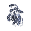 7qjmC  7qjnC  7qjoC  7qjpC  7qjqC  7qjrC  7qjsC  5zoaS S: Starting model for refinement C: citing same article ( |
|---|
-Links
 PDBj
PDBj- Assembly
| Deposited unit | 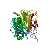
| ||||||||||||
|---|---|---|---|---|---|---|---|---|---|---|---|---|---|
| 1 |
| ||||||||||||
| Unit cell |
| ||||||||||||
| Components on special symmetry positions |
|
-Components
| #1: Protein | Mass: 29146.359 Da / Num. of mol.: 1 Source method: isolated from a genetically manipulated source Source: (gene. exp.) Thermobifida cellulosilytica TB100 (bacteria)Production host: | ||||||
|---|---|---|---|---|---|---|---|
| #2: Chemical | ChemComp-GOL /   Mass: 92.094 Da / Num. of mol.: 1 / Source method: obtained synthetically / Formula: C3H8O3 / Feature type: SUBJECT OF INVESTIGATION Mass: 92.094 Da / Num. of mol.: 1 / Source method: obtained synthetically / Formula: C3H8O3 / Feature type: SUBJECT OF INVESTIGATION | ||||||
| #3: Chemical | ChemComp-PG4 /   Mass: 194.226 Da / Num. of mol.: 1 / Source method: obtained synthetically / Formula: C8H18O5 / Feature type: SUBJECT OF INVESTIGATION / Comment: precipitant*YM Mass: 194.226 Da / Num. of mol.: 1 / Source method: obtained synthetically / Formula: C8H18O5 / Feature type: SUBJECT OF INVESTIGATION / Comment: precipitant*YM | ||||||
| #4: Chemical |   Mass: 24.305 Da / Num. of mol.: 3 / Source method: obtained synthetically / Formula: Mg / Feature type: SUBJECT OF INVESTIGATION Mass: 24.305 Da / Num. of mol.: 3 / Source method: obtained synthetically / Formula: Mg / Feature type: SUBJECT OF INVESTIGATION#5: Water | ChemComp-HOH / |  Mass: 18.015 Da / Num. of mol.: 241 / Source method: isolated from a natural source / Formula: H2O Mass: 18.015 Da / Num. of mol.: 241 / Source method: isolated from a natural source / Formula: H2OHas ligand of interest | Y | Has protein modification | Y | |
-Experimental details
-Experiment
| Experiment | Method: X-RAY DIFFRACTION / Number of used crystals: 1 |
|---|
- Sample preparation
| Crystal | Density Matthews: 2.26 Å3/Da / Density % sol: 45.66 % |
|---|---|
| Crystal grow | Temperature: 293 K / Method: vapor diffusion, sitting drop Details: 0.2 M Magnesium Chloride Hexahydrate, 0.1 M Tris pH 8.5, 20 % (w/v) PEG 8000 |
-Data collection
| Diffraction | Mean temperature: 100 K / Serial crystal experiment: N |
|---|---|
| Diffraction source | Source: SYNCHROTRON / Site: Diamond / Beamline: I03 / Wavelength: 0.9763 Å |
| Detector | Type: DECTRIS EIGER2 XE 16M / Detector: PIXEL / Date: Oct 22, 2020 |
| Radiation | Protocol: SINGLE WAVELENGTH / Monochromatic (M) / Laue (L): M / Scattering type: x-ray |
| Radiation wavelength | Wavelength: 0.9763 Å / Relative weight: 1 |
| Reflection | Resolution: 1.777→77.285 Å / Num. obs: 17061 / % possible obs: 87.1 % / Redundancy: 26.2 % / CC1/2: 0.992 / Rmerge(I) obs: 0.639 / Rpim(I) all: 0.126 / Net I/σ(I): 6.3 |
| Reflection shell | Resolution: 1.777→1.965 Å / Redundancy: 22 % / Rmerge(I) obs: 3.127 / Mean I/σ(I) obs: 1.6 / Num. unique obs: 855 / CC1/2: 0.615 / Rpim(I) all: 0.65 / % possible all: 64.3 |
- Processing
| Software |
| |||||||||||||||||||||||||||||||||||||||||||||||||||||||||||||||||||||||||||||||||||||||||||||||||||||||||||||||||||||||||||||||||||||||||||||||||||||||||||||||||||||||||||||||||||||||||||||||||||||||||||||||||||||||||||||||||||||||
|---|---|---|---|---|---|---|---|---|---|---|---|---|---|---|---|---|---|---|---|---|---|---|---|---|---|---|---|---|---|---|---|---|---|---|---|---|---|---|---|---|---|---|---|---|---|---|---|---|---|---|---|---|---|---|---|---|---|---|---|---|---|---|---|---|---|---|---|---|---|---|---|---|---|---|---|---|---|---|---|---|---|---|---|---|---|---|---|---|---|---|---|---|---|---|---|---|---|---|---|---|---|---|---|---|---|---|---|---|---|---|---|---|---|---|---|---|---|---|---|---|---|---|---|---|---|---|---|---|---|---|---|---|---|---|---|---|---|---|---|---|---|---|---|---|---|---|---|---|---|---|---|---|---|---|---|---|---|---|---|---|---|---|---|---|---|---|---|---|---|---|---|---|---|---|---|---|---|---|---|---|---|---|---|---|---|---|---|---|---|---|---|---|---|---|---|---|---|---|---|---|---|---|---|---|---|---|---|---|---|---|---|---|---|---|---|---|---|---|---|---|---|---|---|---|---|---|---|---|---|---|---|---|
| Refinement | Method to determine structure: MOLECULAR REPLACEMENT Starting model: 5zoa Resolution: 1.78→77.285 Å / Cor.coef. Fo:Fc: 0.952 / Cor.coef. Fo:Fc free: 0.904 / SU B: 8.471 / SU ML: 0.132 / Cross valid method: FREE R-VALUE / ESU R: 0.218 / ESU R Free: 0.19 Details: Hydrogens have been added in their riding positions
| |||||||||||||||||||||||||||||||||||||||||||||||||||||||||||||||||||||||||||||||||||||||||||||||||||||||||||||||||||||||||||||||||||||||||||||||||||||||||||||||||||||||||||||||||||||||||||||||||||||||||||||||||||||||||||||||||||||||
| Solvent computation | Ion probe radii: 0.8 Å / Shrinkage radii: 0.8 Å / VDW probe radii: 1.2 Å / Solvent model: MASK BULK SOLVENT | |||||||||||||||||||||||||||||||||||||||||||||||||||||||||||||||||||||||||||||||||||||||||||||||||||||||||||||||||||||||||||||||||||||||||||||||||||||||||||||||||||||||||||||||||||||||||||||||||||||||||||||||||||||||||||||||||||||||
| Displacement parameters | Biso mean: 18.271 Å2
| |||||||||||||||||||||||||||||||||||||||||||||||||||||||||||||||||||||||||||||||||||||||||||||||||||||||||||||||||||||||||||||||||||||||||||||||||||||||||||||||||||||||||||||||||||||||||||||||||||||||||||||||||||||||||||||||||||||||
| Refinement step | Cycle: LAST / Resolution: 1.78→77.285 Å
| |||||||||||||||||||||||||||||||||||||||||||||||||||||||||||||||||||||||||||||||||||||||||||||||||||||||||||||||||||||||||||||||||||||||||||||||||||||||||||||||||||||||||||||||||||||||||||||||||||||||||||||||||||||||||||||||||||||||
| Refine LS restraints |
| |||||||||||||||||||||||||||||||||||||||||||||||||||||||||||||||||||||||||||||||||||||||||||||||||||||||||||||||||||||||||||||||||||||||||||||||||||||||||||||||||||||||||||||||||||||||||||||||||||||||||||||||||||||||||||||||||||||||
| LS refinement shell | Refine-ID: X-RAY DIFFRACTION / Total num. of bins used: 20
| |||||||||||||||||||||||||||||||||||||||||||||||||||||||||||||||||||||||||||||||||||||||||||||||||||||||||||||||||||||||||||||||||||||||||||||||||||||||||||||||||||||||||||||||||||||||||||||||||||||||||||||||||||||||||||||||||||||||
| Refinement TLS params. | Method: refined / Origin x: -2.8901 Å / Origin y: 26.549 Å / Origin z: -5.7805 Å
| |||||||||||||||||||||||||||||||||||||||||||||||||||||||||||||||||||||||||||||||||||||||||||||||||||||||||||||||||||||||||||||||||||||||||||||||||||||||||||||||||||||||||||||||||||||||||||||||||||||||||||||||||||||||||||||||||||||||
| Refinement TLS group |
|
