 Movie
Movie Controller

[English] 日本語
 Yorodumi
- PDB-7qjj: X-Ray Structure of a Mn2+ soak of EleNRMT in complex with two Nan... -
+ Open data

- Basic information
| Entry | Database: PDB / ID: 7qjj | ||||||
|---|---|---|---|---|---|---|---|
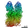
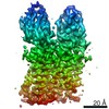
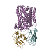
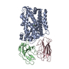
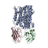
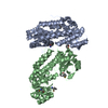
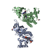
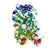
| Title | X-Ray Structure of a Mn2+ soak of EleNRMT in complex with two Nanobodies at 4.6A | ||||||
 Components Components |
| ||||||
 Keywords Keywords | MEMBRANE PROTEIN / SLC11 / NRAMP-related Mg2+ transporter / Nanobody complex | ||||||
| Function / homology |  Function and homology information Function and homology informationmanganese ion transmembrane transporter activity / cadmium ion transmembrane transporter activity / intracellular manganese ion homeostasis / cellular response to iron ion / iron ion transport / plasma membrane Similarity search - Function | ||||||
| Biological species |  | ||||||
| Method |  X-RAY DIFFRACTION / SYNCHROTRON / MOLECULAR REPLACEMENT / Resolution: 4.6 Å X-RAY DIFFRACTION / SYNCHROTRON / MOLECULAR REPLACEMENT / Resolution: 4.6 Å | ||||||
 Authors Authors | Ramanadane, K. / Straub, M.S. / Dutzler, R. / Manatschal, C. | ||||||
| Funding support |  Switzerland, 1items Switzerland, 1items
| ||||||
 Citation Citation | Journal: Elife / Year: 2022 Title: Structural and functional properties of a magnesium transporter of the SLC11/NRAMP family. Authors: Karthik Ramanadane / Monique S Straub / Raimund Dutzler / Cristina Manatschal / Abstract: Members of the ubiquitous SLC11/NRAMP family catalyze the uptake of divalent transition metal ions into cells. They have evolved to efficiently select these trace elements from a large pool of Ca and ...Members of the ubiquitous SLC11/NRAMP family catalyze the uptake of divalent transition metal ions into cells. They have evolved to efficiently select these trace elements from a large pool of Ca and Mg, which are both orders of magnitude more abundant, and to concentrate them in the cytoplasm aided by the cotransport of H serving as energy source. In the present study, we have characterized a member of a distant clade of the family found in prokaryotes, termed NRMTs, that were proposed to function as transporters of Mg. The protein transports Mg and Mn but not Ca by a mechanism that is not coupled to H. Structures determined by cryo-EM and X-ray crystallography revealed a generally similar protein architecture compared to classical NRAMPs, with a restructured ion binding site whose increased volume provides suitable interactions with ions that likely have retained much of their hydration shell. | ||||||
| History |
|
- Structure visualization
| Structure viewer | Molecule: MolmilJmol/JSmol |
|---|
- Downloads & links
-Download
| PDBx/mmCIF format | 7qjj.cif.gz | 601.4 KB | Display | PDBx/mmCIF format |
|---|---|---|---|---|
| PDB format | pdb7qjj.ent.gz | 422.8 KB | Display | PDB format |
| PDBx/mmJSON format | 7qjj.json.gz | Tree view | PDBx/mmJSON format | |
| Others |  Other downloads Other downloads |
-Validation report
| Summary document | 7qjj_validation.pdf.gz | 452.5 KB | Display | wwPDB validaton report |
|---|---|---|---|---|
| Full document | 7qjj_full_validation.pdf.gz | 471.4 KB | Display | |
| Data in XML | 7qjj_validation.xml.gz | 45.2 KB | Display | |
| Data in CIF | 7qjj_validation.cif.gz | 61.1 KB | Display | |
| Arichive directory | https://data.pdbj.org/pub/pdb/validation_reports/qj/7qjjftp://data.pdbj.org/pub/pdb/validation_reports/qj/7qjj | HTTPS FTP |
-Related structure data
| Related structure data | 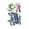 7qiaSC  7qicC  7qjiC C: citing same article ( S: Starting model for refinement |
|---|---|
| Similar structure data |


-Links
 PDBj
PDBj

- Assembly
| Deposited unit | 
| ||||||||||||||||||||||||||||||||||||||||||||||||||||||||||||||||||||||||||||||||||||||||||
|---|---|---|---|---|---|---|---|---|---|---|---|---|---|---|---|---|---|---|---|---|---|---|---|---|---|---|---|---|---|---|---|---|---|---|---|---|---|---|---|---|---|---|---|---|---|---|---|---|---|---|---|---|---|---|---|---|---|---|---|---|---|---|---|---|---|---|---|---|---|---|---|---|---|---|---|---|---|---|---|---|---|---|---|---|---|---|---|---|---|---|---|
| 1 | 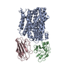
| ||||||||||||||||||||||||||||||||||||||||||||||||||||||||||||||||||||||||||||||||||||||||||
| 2 | 
| ||||||||||||||||||||||||||||||||||||||||||||||||||||||||||||||||||||||||||||||||||||||||||
| Unit cell |
| ||||||||||||||||||||||||||||||||||||||||||||||||||||||||||||||||||||||||||||||||||||||||||
| Noncrystallographic symmetry (NCS) | NCS domain:
NCS domain segments:
NCS ensembles :
NCS oper:
|
-Components
| #1: Protein | Mass: 46848.828 Da / Num. of mol.: 2 Source method: isolated from a genetically manipulated source Source: (gene. exp.)  #2: Antibody | Mass: 12952.605 Da / Num. of mol.: 2 Source method: isolated from a genetically manipulated source Source: (gene. exp.) #3: Antibody | Mass: 13351.896 Da / Num. of mol.: 2 Source method: isolated from a genetically manipulated source Source: (gene. exp.) #4: Chemical |   Mass: 54.938 Da / Num. of mol.: 2 / Source method: obtained synthetically / Formula: Mn Mass: 54.938 Da / Num. of mol.: 2 / Source method: obtained synthetically / Formula: MnHas ligand of interest | N | |
|---|
-Experimental details
-Experiment
| Experiment | Method: X-RAY DIFFRACTION / Number of used crystals: 1 |
|---|
- Sample preparation
| Crystal | Density Matthews: 2.59 Å3/Da / Density % sol: 52.49 % |
|---|---|
| Crystal grow | Temperature: 277.17 K / Method: vapor diffusion, sitting drop Details: 50 mM MgAc, 50 mM HEPES pH 7.2-7.6 and 25-30% PEG400 and crystals soaked in 25mM MnAc |
-Data collection
| Diffraction | Mean temperature: 80 K / Serial crystal experiment: N |
|---|---|
| Diffraction source | Source: SYNCHROTRON / Site: SLS / Beamline: X06DA / Wavelength: 1.892738 Å |
| Detector | Type: DECTRIS PILATUS 2M-F / Detector: PIXEL / Date: Mar 27, 2021 |
| Radiation | Protocol: SINGLE WAVELENGTH / Monochromatic (M) / Laue (L): M / Scattering type: x-ray |
| Radiation wavelength | Wavelength: 1.892738 Å / Relative weight: 1 |
| Reflection | Resolution: 4.6→12 Å / Num. obs: 32816 / % possible obs: 99.7 % / Redundancy: 14 % / Biso Wilson estimate: 189.78 Å2 / CC1/2: 1 / Rrim(I) all: 0.144 / Net I/σ(I): 11.42 |
| Reflection shell | Resolution: 4.6→4.7 Å / Mean I/σ(I) obs: 1.85 / Num. unique obs: 2039 / CC1/2: 0.91 / Rrim(I) all: 1.474 |
- Processing
| Software |
| |||||||||||||||||||||||||||||||||||||||||||||||||
|---|---|---|---|---|---|---|---|---|---|---|---|---|---|---|---|---|---|---|---|---|---|---|---|---|---|---|---|---|---|---|---|---|---|---|---|---|---|---|---|---|---|---|---|---|---|---|---|---|---|---|
| Refinement | Method to determine structure: MOLECULAR REPLACEMENT Starting model: PDBID 7QIA Resolution: 4.6→12 Å / SU ML: 0.7298 / Cross valid method: FREE R-VALUE / σ(F): 1.37 / Phase error: 40.4518 Stereochemistry target values: GeoStd + Monomer Library + CDL v1.2
| |||||||||||||||||||||||||||||||||||||||||||||||||
| Solvent computation | Shrinkage radii: 0.9 Å / VDW probe radii: 1.1 Å / Solvent model: FLAT BULK SOLVENT MODEL | |||||||||||||||||||||||||||||||||||||||||||||||||
| Displacement parameters | Biso mean: 270.79 Å2 | |||||||||||||||||||||||||||||||||||||||||||||||||
| Refinement step | Cycle: LAST / Resolution: 4.6→12 Å
| |||||||||||||||||||||||||||||||||||||||||||||||||
| Refine LS restraints |
| |||||||||||||||||||||||||||||||||||||||||||||||||
| Refine LS restraints NCS |
| |||||||||||||||||||||||||||||||||||||||||||||||||
| LS refinement shell |
| |||||||||||||||||||||||||||||||||||||||||||||||||
| Refinement TLS params. | Method: refined / Origin x: -23.375792412 Å / Origin y: -20.0962169733 Å / Origin z: -30.5290581484 Å
| |||||||||||||||||||||||||||||||||||||||||||||||||
| Refinement TLS group | Selection details: all |
