 Movie
Movie Controller

[English] 日本語
 Yorodumi
- PDB-7qf3: Structure of the R57Q mutant of miniSOG expressed in E. coli in r... -
+ Open data

- Basic information
| Entry | Database: PDB / ID: 7qf3 | ||||||
|---|---|---|---|---|---|---|---|
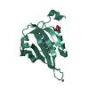
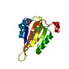
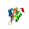
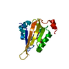
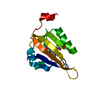
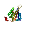
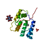
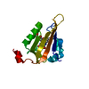
| Title | Structure of the R57Q mutant of miniSOG expressed in E. coli in regular LB medium | ||||||
 Components Components | miniSOG (R57Q mutant) | ||||||
 Keywords Keywords | FLAVOPROTEIN / LOV DOMAIN / RIBOFLAVIN / PHOTOSENSITIZING PROTEIN / FLUORESCENT PROTEIN | ||||||
| Function / homology | : / FLAVIN MONONUCLEOTIDE Function and homology information Function and homology information | ||||||
| Biological species |  | ||||||
| Method |  X-RAY DIFFRACTION / SYNCHROTRON / MOLECULAR REPLACEMENT / Resolution: 1.1 Å X-RAY DIFFRACTION / SYNCHROTRON / MOLECULAR REPLACEMENT / Resolution: 1.1 Å | ||||||
 Authors Authors | Lafaye, C. / Aumonier, S. / von Stetten, D. / Noirclerc-Savoye, N. / Gotthard, G. / Royant, A. | ||||||
| Funding support |  France, 1items France, 1items
| ||||||
 Citation Citation | Journal: Photochem Photobiol Sci / Year: 2022 Title: Riboflavin-binding proteins for singlet oxygen production. Authors: Lafaye, C. / Aumonier, S. / Torra, J. / Signor, L. / von Stetten, D. / Noirclerc-Savoye, M. / Shu, X. / Ruiz-Gonzalez, R. / Gotthard, G. / Royant, A. / Nonell, S. #1: Journal: Sci Rep / Year: 2019Title: Tailing miniSOG: structural bases of the complex photophysics of a flavin-binding singlet oxygen photosensitizing protein. Authors: Torra, J. / Lafaye, C. / Signor, L. / Aumonier, S. / Flors, C. / Shu, X. / Nonell, S. / Gotthard, G. / Royant, A. | ||||||
| History |
|
- Structure visualization
| Structure viewer | Molecule: MolmilJmol/JSmol |
|---|
- Downloads & links
-Download
| PDBx/mmCIF format | 7qf3.cif.gz | 71.1 KB | Display | PDBx/mmCIF format |
|---|---|---|---|---|
| PDB format | pdb7qf3.ent.gz | Display | PDB format | |
| PDBx/mmJSON format | 7qf3.json.gz | Tree view | PDBx/mmJSON format | |
| Others |  Other downloads Other downloads |
-Validation report
| Arichive directory | https://data.pdbj.org/pub/pdb/validation_reports/qf/7qf3ftp://data.pdbj.org/pub/pdb/validation_reports/qf/7qf3 | HTTPS FTP |
|---|
-Related structure data
| Related structure data |  7qf2C 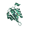 7qf4C  7qf5C  6gpuS S: Starting model for refinement C: citing same article ( |
|---|---|
| Similar structure data |


-Links
 PDBj
PDBj
- Assembly
| Deposited unit | 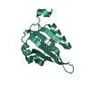
| ||||||||
|---|---|---|---|---|---|---|---|---|---|
| 1 |
| ||||||||
| Unit cell |
|
-Components
-Protein , 1 types, 1 molecules AAA
| #1: Protein | Mass: 14855.712 Da / Num. of mol.: 1 Source method: isolated from a genetically manipulated source Source: (gene. exp.) Production host:  |
|---|
-Non-polymers , 5 types, 104 molecules 


| #2: Chemical | ChemComp-FMN /  Mass: 456.344 Da / Num. of mol.: 1 / Source method: obtained synthetically / Formula: C17H21N4O9P / Feature type: SUBJECT OF INVESTIGATION Mass: 456.344 Da / Num. of mol.: 1 / Source method: obtained synthetically / Formula: C17H21N4O9P / Feature type: SUBJECT OF INVESTIGATION |
|---|---|
| #3: Chemical | ChemComp-MG /  Mass: 24.305 Da / Num. of mol.: 1 / Source method: obtained synthetically / Formula: Mg Mass: 24.305 Da / Num. of mol.: 1 / Source method: obtained synthetically / Formula: Mg |
| #4: Chemical | ChemComp-CO /  Mass: 58.933 Da / Num. of mol.: 1 / Source method: obtained synthetically / Formula: Co Mass: 58.933 Da / Num. of mol.: 1 / Source method: obtained synthetically / Formula: Co |
| #5: Chemical | ChemComp-CL /  Mass: 35.453 Da / Num. of mol.: 1 / Source method: obtained synthetically / Formula: Cl Mass: 35.453 Da / Num. of mol.: 1 / Source method: obtained synthetically / Formula: Cl |
| #6: Water | ChemComp-HOH / Mass: 18.015 Da / Num. of mol.: 100 / Source method: isolated from a natural source / Formula: H2O |
-Details
| Has ligand of interest | Y |
|---|
-Experimental details
-Experiment
| Experiment | Method: X-RAY DIFFRACTION / Number of used crystals: 1 |
|---|
- Sample preparation
| Crystal | Density Matthews: 2.04 Å3/Da / Density % sol: 39.8 % |
|---|---|
| Crystal grow | Temperature: 293 K / Method: vapor diffusion, hanging drop / pH: 8 Details: 100 mM Tris-Hcl pH 8.0, 20 mM MgCl2, 28% PEG 4000, 15 mM CoCl2 |
-Data collection
| Diffraction | Mean temperature: 100 K / Serial crystal experiment: N |
|---|---|
| Diffraction source | Source: SYNCHROTRON / Site: ESRF / Beamline: ID29 / Wavelength: 0.9322 Å |
| Detector | Type: DECTRIS PILATUS3 6M / Detector: PIXEL / Date: May 21, 2014 / Details: TOROIDAL MIRROR |
| Radiation | Monochromator: Si(111) / Protocol: SINGLE WAVELENGTH / Monochromatic (M) / Laue (L): M / Scattering type: x-ray |
| Radiation wavelength | Wavelength: 0.9322 Å / Relative weight: 1 |
| Reflection | Resolution: 1.1→38.28 Å / Num. obs: 45101 / % possible obs: 100 % / Redundancy: 12.4 % / Biso Wilson estimate: 14.5 Å2 / CC1/2: 1 / Rrim(I) all: 0.084 / Net I/σ(I): 15.5 |
| Reflection shell | Resolution: 1.1→1.14 Å / Redundancy: 11.9 % / Mean I/σ(I) obs: 1.9 / Num. unique obs: 4488 / CC1/2: 0.596 / Rrim(I) all: 1.199 / % possible all: 100 |
- Processing
| Software |
| |||||||||||||||||||||||||||||||||||||||||||||||||||||||||||||||||||||||||||||||||||||||||||||||||||||||||||||||||||||||||||||||||||||||||||||||||||||||||||||||||||||
|---|---|---|---|---|---|---|---|---|---|---|---|---|---|---|---|---|---|---|---|---|---|---|---|---|---|---|---|---|---|---|---|---|---|---|---|---|---|---|---|---|---|---|---|---|---|---|---|---|---|---|---|---|---|---|---|---|---|---|---|---|---|---|---|---|---|---|---|---|---|---|---|---|---|---|---|---|---|---|---|---|---|---|---|---|---|---|---|---|---|---|---|---|---|---|---|---|---|---|---|---|---|---|---|---|---|---|---|---|---|---|---|---|---|---|---|---|---|---|---|---|---|---|---|---|---|---|---|---|---|---|---|---|---|---|---|---|---|---|---|---|---|---|---|---|---|---|---|---|---|---|---|---|---|---|---|---|---|---|---|---|---|---|---|---|---|---|
| Refinement | Method to determine structure: MOLECULAR REPLACEMENT Starting model: 6GPU Resolution: 1.1→38.28 Å / Cor.coef. Fo:Fc: 0.974 / Cor.coef. Fo:Fc free: 0.973 / SU B: 1.169 / SU ML: 0.024 / Cross valid method: FREE R-VALUE / ESU R: 0.031 / ESU R Free: 0.032 Details: Hydrogens have been added in their riding positions
| |||||||||||||||||||||||||||||||||||||||||||||||||||||||||||||||||||||||||||||||||||||||||||||||||||||||||||||||||||||||||||||||||||||||||||||||||||||||||||||||||||||
| Solvent computation | Ion probe radii: 0.8 Å / Shrinkage radii: 0.8 Å / VDW probe radii: 1.2 Å / Solvent model: MASK BULK SOLVENT | |||||||||||||||||||||||||||||||||||||||||||||||||||||||||||||||||||||||||||||||||||||||||||||||||||||||||||||||||||||||||||||||||||||||||||||||||||||||||||||||||||||
| Displacement parameters | Biso mean: 13.784 Å2
| |||||||||||||||||||||||||||||||||||||||||||||||||||||||||||||||||||||||||||||||||||||||||||||||||||||||||||||||||||||||||||||||||||||||||||||||||||||||||||||||||||||
| Refinement step | Cycle: LAST / Resolution: 1.1→38.28 Å
| |||||||||||||||||||||||||||||||||||||||||||||||||||||||||||||||||||||||||||||||||||||||||||||||||||||||||||||||||||||||||||||||||||||||||||||||||||||||||||||||||||||
| Refine LS restraints |
| |||||||||||||||||||||||||||||||||||||||||||||||||||||||||||||||||||||||||||||||||||||||||||||||||||||||||||||||||||||||||||||||||||||||||||||||||||||||||||||||||||||
| LS refinement shell |
|
