 Movie
Movie Controller

[English] 日本語
 Yorodumi
- PDB-7qea: Crystal structure of fluorescein-di-Beta-D-glucuronide bound to a... -
+ Open data

- Basic information
| Entry | Database: PDB / ID: 7qea | ||||||
|---|---|---|---|---|---|---|---|
| Title | Crystal structure of fluorescein-di-Beta-D-glucuronide bound to a mutant of SN243 (D415A) | ||||||
 Components Components | SN243 | ||||||
 Keywords Keywords | HYDROLASE / enzyme discovery / carbohydrate-active enzymes (CAZy) / protein engineering / functional metagenomics) | ||||||
| Function / homology | ACETATE ION / Chem-B9I Function and homology information Function and homology information | ||||||
| Biological species | Synthetic construct (others) | ||||||
| Method |  X-RAY DIFFRACTION / SYNCHROTRON / MOLECULAR REPLACEMENT / Resolution: 2.28 Å X-RAY DIFFRACTION / SYNCHROTRON / MOLECULAR REPLACEMENT / Resolution: 2.28 Å | ||||||
 Authors Authors | Neun, S. / Brear, P. / Campbell, E. / Omari, K. / Wagner, O. / Hyvonen, M. / Hollfelder, F. | ||||||
| Funding support | European Union, 1items
| ||||||
 Citation Citation | Journal: Nat.Chem.Biol. / Year: 2022 Title: Functional metagenomic screening identifies an unexpected beta-glucuronidase. Authors: Neun, S. / Brear, P. / Campbell, E. / Tryfona, T. / El Omari, K. / Wagner, A. / Dupree, P. / Hyvonen, M. / Hollfelder, F. | ||||||
| History |
|
- Structure visualization
| Structure viewer | Molecule: MolmilJmol/JSmol |
|---|
- Downloads & links
-Download
| PDBx/mmCIF format | 7qea.cif.gz | 304.9 KB | Display | PDBx/mmCIF format |
|---|---|---|---|---|
| PDB format | pdb7qea.ent.gz | 241.1 KB | Display | PDB format |
| PDBx/mmJSON format | 7qea.json.gz | Tree view | PDBx/mmJSON format | |
| Others |  Other downloads Other downloads |
-Validation report
| Arichive directory | https://data.pdbj.org/pub/pdb/validation_reports/qe/7qeaftp://data.pdbj.org/pub/pdb/validation_reports/qe/7qea | HTTPS FTP |
|---|
-Related structure data
| Related structure data | 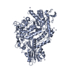 7qe1SC  7qe2C  7qeeC 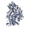 7qefC 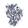 7qg4C S: Starting model for refinement C: citing same article ( |
|---|---|
| Similar structure data |
-Links
 PDBj
PDBj- Assembly
| Deposited unit | 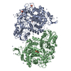
| |||||||||||||||||||||||||||||||||||||||||||||||||||||||
|---|---|---|---|---|---|---|---|---|---|---|---|---|---|---|---|---|---|---|---|---|---|---|---|---|---|---|---|---|---|---|---|---|---|---|---|---|---|---|---|---|---|---|---|---|---|---|---|---|---|---|---|---|---|---|---|---|
| 1 | 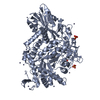
| |||||||||||||||||||||||||||||||||||||||||||||||||||||||
| 2 | 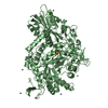
| |||||||||||||||||||||||||||||||||||||||||||||||||||||||
| Unit cell |
| |||||||||||||||||||||||||||||||||||||||||||||||||||||||
| Noncrystallographic symmetry (NCS) | NCS domain:
NCS domain segments:
|