| 登録情報 | データベース: PDB / ID: 7q5i
|
|---|
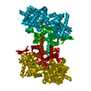
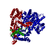
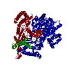
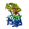
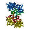
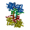
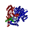
| タイトル | A glucose-based molecular rotor probes the catalytic site of glycogen phosphorylase. |
|---|
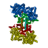
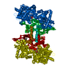
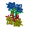
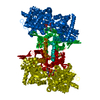
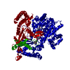
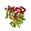
 要素 要素 | Glycogen phosphorylase, muscle form |
|---|
 キーワード キーワード | SUGAR BINDING PROTEIN / Glycogen phosphorylase / inhibitor / type 2 diabetes / molecular rotors |
|---|
| 機能・相同性 |  機能・相同性情報 機能・相同性情報
glycogen phosphorylase / glycogen phosphorylase activity / glycogen catabolic process / skeletal muscle myofibril / pyridoxal phosphate binding / nucleotide binding類似検索 - 分子機能 Glycosyl transferase, family 35 / Glycogen/starch/alpha-glucan phosphorylase / Phosphorylase pyridoxal-phosphate attachment site / Carbohydrate phosphorylase / Phosphorylase pyridoxal-phosphate attachment site.類似検索 - ドメイン・相同性 BETA-MERCAPTOETHANOL / CARBONATE ION / Chem-I0F / Glycogen phosphorylase, muscle form類似検索 - 構成要素 |
|---|
| 生物種 |   Oryctolagus cuniculus (ウサギ) Oryctolagus cuniculus (ウサギ) |
|---|
| 手法 |  X線回折 / シンクロトロン / 分子置換 / 解像度: 1.8 Å X線回折 / シンクロトロン / 分子置換 / 解像度: 1.8 Å |
|---|
 データ登録者 データ登録者 | Neofytos, D.D. / Chrysina, E.D. |
|---|
| 資金援助 |  ギリシャ, European Union, 3件 ギリシャ, European Union, 3件 | 組織 | 認可番号 | 国 |
|---|
| Other government | MIS 5002550 | ギリシャ | | European Commission | Project ID: 653706 | European Union | | Other government | MIS 5048135 | ギリシャ |
|
|---|
 引用 引用 | ジャーナル: Org.Biomol.Chem. / 年: 2022タイトル: A glucose-based molecular rotor inhibitor of glycogen phosphorylase as a probe of cellular enzymatic function. 著者: Minadakis, M.P. / Mavreas, K.F. / Neofytos, D.D. / Paschou, M. / Kogkaki, A. / Athanasiou, V. / Mamais, M. / Veclani, D. / Iatrou, H. / Venturini, A. / Chrysina, E.D. / Papazafiri, P. / Gimisis, T. |
|---|
| 履歴 | | 登録 | 2021年11月3日 | 登録サイト: PDBE / 処理サイト: PDBE |
|---|
| 改定 1.0 | 2022年3月2日 | Provider: repository / タイプ: Initial release |
|---|
| 改定 2.0 | 2022年3月9日 | Group: Advisory / Atomic model ...Advisory / Atomic model / Data collection / Non-polymer description / Structure summary
カテゴリ: atom_site / chem_comp ...atom_site / chem_comp / database_PDB_caveat / entity / pdbx_validate_chiral
Item: _atom_site.auth_atom_id / _atom_site.label_atom_id ..._atom_site.auth_atom_id / _atom_site.label_atom_id / _chem_comp.formula / _chem_comp.formula_weight / _entity.formula_weight |
|---|
| 改定 2.1 | 2022年3月30日 | Group: Database references / カテゴリ: citation
Item: _citation.journal_volume / _citation.page_first / _citation.page_last |
|---|
| 改定 2.2 | 2024年1月31日 | Group: Data collection / Derived calculations / Refinement description
カテゴリ: atom_type / chem_comp_atom ...atom_type / chem_comp_atom / chem_comp_bond / pdbx_initial_refinement_model
Item: _atom_type.pdbx_N_electrons / _atom_type.pdbx_scat_Z |
|---|
|
|---|
 ムービー
ムービー コントローラー


 データを開く
 基本情報
基本情報 構造の表示
構造の表示 ダウンロードとリンク
 その他のダウンロード
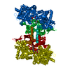


 PDBj
PDBj


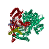
 集合体
集合体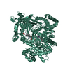

 分子量: 377.392 Da / 分子数: 1 / 由来タイプ: 合成 / 式: C18H23N3O6 / タイプ: SUBJECT OF INVESTIGATION
 分子量: 60.009 Da / 分子数: 3 / 由来タイプ: 合成 / 式: CO3
 分子量: 78.133 Da / 分子数: 1 / 由来タイプ: 合成 / 式: C2H6OS
 分子量: 18.015 Da / 分子数: 251 / 由来タイプ: 天然 / 式: H2O
 試料調製
試料調製 / ビームライン: P13 (MX1) / 波長: 0.9763 Å
 解析
解析