 Movie
Movie Controller

[English] 日本語
 Yorodumi
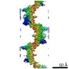
- PDB-7q3n: Cryo-EM of the complex between human uromodulin (UMOD)/Tamm-Horsf... -
+ Open data

- Basic information
| Entry | Database: PDB / ID: 7q3n | |||||||||||||||
|---|---|---|---|---|---|---|---|---|---|---|---|---|---|---|---|---|
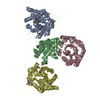
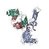
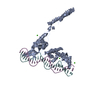
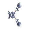
| Title | Cryo-EM of the complex between human uromodulin (UMOD)/Tamm-Horsfall protein (THP) and the FimH lectin domain from uropathogenic E. coli | |||||||||||||||
 Components Components |
| |||||||||||||||
 Keywords Keywords | ANTIMICROBIAL PROTEIN / EGF DOMAIN / DECOY MODULE / BETA-HAIRPIN / D10C DOMAIN / D8C DOMAIN / EXTRACELLULAR MATRIX / GLYCOPROTEIN / N-GLYCAN / HIGH-MANNOSE SUGAR / CELL ADHESION / BACTERIAL ADHESIN / TYPE I PILUS / SUGAR BINDING PROTEIN / LECTIN / URINARY TRACT INFECTION / UTI / UROPATHOGENIC E. COLI / UPEC | |||||||||||||||
| Function / homology |  Function and homology information Function and homology informationcitric acid secretion / protein localization to extracellular region / metanephric thick ascending limb development / metanephric distal convoluted tubule development / connective tissue replacement / protein transport into plasma membrane raft / Asparagine N-linked glycosylation / organ or tissue specific immune response / collecting duct development / urea transmembrane transport ...citric acid secretion / protein localization to extracellular region / metanephric thick ascending limb development / metanephric distal convoluted tubule development / connective tissue replacement / protein transport into plasma membrane raft / Asparagine N-linked glycosylation / organ or tissue specific immune response / collecting duct development / urea transmembrane transport / micturition / metanephric ascending thin limb development / protein localization to vacuole / regulation of protein transport / juxtaglomerular apparatus development / intracellular chloride ion homeostasis / renal urate salt excretion / glomerular filtration / urate transport / antibacterial innate immune response / renal sodium ion absorption / neutrophil migration / response to water deprivation / regulation of urine volume / potassium ion homeostasis / intracellular sodium ion homeostasis / endoplasmic reticulum organization / intracellular phosphate ion homeostasis / cell adhesion involved in single-species biofilm formation / heterophilic cell-cell adhesion / IgG binding / pilus / extrinsic component of membrane / leukocyte cell-cell adhesion / ciliary membrane / cellular response to unfolded protein / multicellular organismal response to stress / cellular defense response / renal water homeostasis / side of membrane / ERAD pathway / RNA splicing / tumor necrosis factor-mediated signaling pathway / lipid metabolic process / apoptotic signaling pathway / Golgi lumen / regulation of blood pressure / autophagy / intracellular calcium ion homeostasis / spindle pole / protein folding / response to lipopolysaccharide / gene expression / defense response to Gram-negative bacterium / basolateral plasma membrane / apical plasma membrane / cilium / response to xenobiotic stimulus / inflammatory response / negative regulation of cell population proliferation / calcium ion binding / cell surface / endoplasmic reticulum / : / extracellular exosome / membrane Similarity search - Function | |||||||||||||||
| Biological species |   Homo sapiens (human) Homo sapiens (human) | |||||||||||||||
| Method | ELECTRON MICROSCOPY / single particle reconstruction / cryo EM / Resolution: 7.4 Å | |||||||||||||||
 Authors Authors | Jovine, L. / Xu, C. / Stsiapanava, A. / Carroni, M. / Tunyasuvunakool, K. / Jumper, J. / Wu, B. | |||||||||||||||
| Funding support |  Singapore, 4items Singapore, 4items
| |||||||||||||||
 Citation Citation | Journal: Nat Struct Mol Biol / Year: 2022 Title: Structure of the decoy module of human glycoprotein 2 and uromodulin and its interaction with bacterial adhesin FimH. Authors: Alena Stsiapanava / Chenrui Xu / Shunsuke Nishio / Ling Han / Nao Yamakawa / Marta Carroni / Kathryn Tunyasuvunakool / John Jumper / Daniele de Sanctis / Bin Wu / Luca Jovine /    Abstract: Glycoprotein 2 (GP2) and uromodulin (UMOD) filaments protect against gastrointestinal and urinary tract infections by acting as decoys for bacterial fimbrial lectin FimH. By combining AlphaFold2 ...Glycoprotein 2 (GP2) and uromodulin (UMOD) filaments protect against gastrointestinal and urinary tract infections by acting as decoys for bacterial fimbrial lectin FimH. By combining AlphaFold2 predictions with X-ray crystallography and cryo-EM, we show that these proteins contain a bipartite decoy module whose new fold presents the high-mannose glycan recognized by FimH. The structure rationalizes UMOD mutations associated with kidney diseases and visualizes a key epitope implicated in cast nephropathy. #1: Journal: Proc Soc Exp Biol Med / Year: 1950 Title: Characterization and separation of an inhibitor of viral hemagglutination present in urine. #2: Journal: Science / Year: 1987 Title: Identification of human uromodulin as the Tamm-Horsfall urinary glycoprotein. Abstract: The primary structure of human uromodulin, a 616-amino acid, 85-kilodalton glycoprotein with in vitro immunosuppressive properties, was determined through isolation and characterization of ...The primary structure of human uromodulin, a 616-amino acid, 85-kilodalton glycoprotein with in vitro immunosuppressive properties, was determined through isolation and characterization of complementary DNA and genomic clones. The amino acid sequence encoded by one of the exons of the uromodulin gene has homology to the low-density-lipoprotein receptor and the epidermal growth factor precursor. Northern hybridization analyses demonstrate that uromodulin is synthesized by the kidney. Evidence is provided that uromodulin is identical to the previously characterized Tamm-Horsfall glycoprotein, the most abundant protein in normal human urine. #3: Journal: J Biol Chem / Year: 2001 Title: Tamm-Horsfall protein binds to type 1 fimbriated Escherichia coli and prevents E. coli from binding to uroplakin Ia and Ib receptors. Authors: J Pak / Y Pu / Z T Zhang / D L Hasty / X R Wu /  Abstract: The adherence of uropathogenic Escherichia coli to the urothelial surface, a critical first step in the pathogenesis of urinary tract infection (UTI), is controlled by three key elements: E. coli ...The adherence of uropathogenic Escherichia coli to the urothelial surface, a critical first step in the pathogenesis of urinary tract infection (UTI), is controlled by three key elements: E. coli adhesins, host receptors, and host defense mechanisms. Although much has been learned about E. coli adhesins and their urothelial receptors, little is known about the role of host defense in the adherence process. Here we show that Tamm-Horsfall protein (THP) is the principal urinary protein that binds specifically to type 1 fimbriated E. coli, the main cause of UTI. The binding was highly specific and saturable and could be inhibited by d-mannose and abolished by endoglycosidase H treatment of THP, suggesting that the binding is mediated by the high-mannose moieties of THP. It is species-conserved, occurring in both human and mouse THPs. In addition, the binding to THP was much greater with an E. coli strain bearing a phenotypic variant of the type 1 fimbrial FimH adhesin characteristic of those prevalent in UTI isolates compared with the one prevalent in isolates from the large intestine of healthy individuals. Finally, a physiological concentration of THP completely abolished the binding of type 1 fimbriated E. coli to uroplakins Ia and Ib, two putative urothelial receptors for type 1 fimbriae. These results establish, on a functional level, that THP contains conserved high-mannose moieties capable of specific interaction with type 1 fimbriae and strongly suggest that this major urinary glycoprotein is a key urinary anti-adherence factor serving to prevent type 1 fimbriated E. coli from binding to the urothelial receptors. #4: Journal: Am J Kidney Dis / Year: 2003 Title: Tamm-Horsfall glycoprotein: biology and clinical relevance. Authors: Franca Serafini-Cessi / Nadia Malagolini / Daniela Cavallone /  Abstract: Tamm-Horsfall glycoprotein (THP) is the most abundant urinary protein in mammals. Urinary excretion occurs by proteolytic cleavage of the large ectodomain of the glycosyl phosphatidylinositol- ...Tamm-Horsfall glycoprotein (THP) is the most abundant urinary protein in mammals. Urinary excretion occurs by proteolytic cleavage of the large ectodomain of the glycosyl phosphatidylinositol-anchored counterpart exposed at the luminal cell surface of the thick ascending limb of Henle's loop. We describe the physical-chemical structure of human THP and its biosynthesis and interaction with other proteins and leukocytes. The clinical relevance of THP reported here includes: (1) involvement in the pathogenesis of cast nephropathy, urolithiasis, and tubulointerstitial nephritis; (2) abnormalities in urinary excretion in renal diseases; and (3) the recent finding that familial juvenile hyperuricemic nephropathy and autosomal dominant medullary cystic kidney disease 2 arise from mutations of the THP gene. We critically examine the literature on the physiological role and mechanism(s) that promote urinary excretion of THP. Some lines of research deal with the in vitro immunoregulatory activity of THP, termed uromodulin when isolated from urine of pregnant women. However, an immunoregulatory function in vivo has not yet been established. In the most recent literature, there is renewed interest in the capacity of urinary THP to compete efficiently with urothelial cell receptors, such as uroplakins, in adhering to type 1 fimbriated Escherichia coli. This property supports the notion that abundant THP excretion in urine is promoted in the host by selective pressure to obtain an efficient defense against urinary tract infections caused by uropathogenic bacteria. #5: Journal: FEBS Lett / Year: 2004 Title: Identification and characterization of D8C, a novel domain present in liver-specific LZP, uromodulin and glycoprotein 2, mutated in familial juvenile hyperuricaemic nephropathy. Authors: Huirong Yang / Chaoqun Wu / Shouyuan Zhao / Jinhu Guo /  Abstract: Present work reported a novel domain--D8C (domain with conserved eight cysteines in liver-specific ZP domain-containing protein, glycoprotein 2 (GP-2) and uromodulin (UMOD)), present in liver- ...Present work reported a novel domain--D8C (domain with conserved eight cysteines in liver-specific ZP domain-containing protein, glycoprotein 2 (GP-2) and uromodulin (UMOD)), present in liver-specific LZP, UMOD, GP-2 and some uncharacterized proteins, most of which are membrane proteins, extracellular proteins or nuclear membrane proteins. D8C contains eight well-conserved cysteine residues, which were predicted to form four pairs of disulfide bridges. D8C is composed mainly of beta-strands. Mutation in the D8C at Cys217 in human UMOD is associated with familial juvenile hyperuricaemic nephropathy, which might be due to the disruption of the disulfide bridge. Identification of D8C would further the understandings of related proteins. #6: Journal: Sci Adv / Year: 2017Title: Evolutionary fine-tuning of conformational ensembles in FimH during host-pathogen interactions. Authors: Vasilios Kalas / Jerome S Pinkner / Thomas J Hannan / Michael E Hibbing / Karen W Dodson / Alex S Holehouse / Hao Zhang / Niraj H Tolia / Michael L Gross / Rohit V Pappu / James Janetka / Scott J Hultgren / Abstract: Positive selection in the two-domain type 1 pilus adhesin FimH enhances fitness in urinary tract infection (UTI). We report a comprehensive atomic-level view of FimH in two-state conformational ...Positive selection in the two-domain type 1 pilus adhesin FimH enhances fitness in urinary tract infection (UTI). We report a comprehensive atomic-level view of FimH in two-state conformational ensembles in solution, composed of one low-affinity tense (T) and multiple high-affinity relaxed (R) conformations. Positively selected residues allosterically modulate the equilibrium between these two conformational states, each of which engages mannose through distinct binding orientations. A FimH variant that only adopts the R state is severely attenuated early in a mouse model of uncomplicated UTI but is proficient at colonizing catheterized bladders in vivo or bladder transitional-like epithelial cells in vitro. Thus, the bladder habitat has barrier(s) to R state-mediated colonization possibly conferred by the terminally differentiated bladder epithelium and/or decoy receptors in urine. Together, our studies reveal the conformational landscape in solution, binding mechanisms, and adhesive strength of an allosteric two-domain adhesin that evolved "moderate" affinity to optimize persistence in the bladder during UTI. #7: Journal: J Am Chem Soc / Year: 2019Title: Binding of the Bacterial Adhesin FimH to Its Natural, Multivalent High-Mannose Type Glycan Targets. Authors: Maximilian M Sauer / Roman P Jakob / Thomas Luber / Fabia Canonica / Giulio Navarra / Beat Ernst / Carlo Unverzagt / Timm Maier / Rudi Glockshuber /   Abstract: Multivalent carbohydrate-lectin interactions at host-pathogen interfaces play a crucial role in the establishment of infections. Although competitive antagonists that prevent pathogen adhesion are ...Multivalent carbohydrate-lectin interactions at host-pathogen interfaces play a crucial role in the establishment of infections. Although competitive antagonists that prevent pathogen adhesion are promising antimicrobial drugs, the molecular mechanisms underlying these complex adhesion processes are still poorly understood. Here, we characterize the interactions between the fimbrial adhesin FimH from uropathogenic Escherichia coli strains and its natural high-mannose type N-glycan binding epitopes on uroepithelial glycoproteins. Crystal structures and a detailed kinetic characterization of ligand-binding and dissociation revealed that the binding pocket of FimH evolved such that it recognizes the terminal α(1-2)-, α(1-3)-, and α(1-6)-linked mannosides of natural high-mannose type N-glycans with similar affinity. We demonstrate that the 2000-fold higher affinity of the domain-separated state of FimH compared to its domain-associated state is ligand-independent and consistent with a thermodynamic cycle in which ligand-binding shifts the association equilibrium between the FimH lectin and the FimH pilin domain. Moreover, we show that a single N-glycan can bind up to three molecules of FimH, albeit with negative cooperativity, so that a molar excess of accessible N-glycans over FimH on the cell surface favors monovalent FimH binding. Our data provide pivotal insights into the adhesion properties of uropathogenic Escherichia coli strains to their target receptors and a solid basis for the development of effective FimH antagonists. #8: Journal: Science / Year: 2020Title: Architecture and function of human uromodulin filaments in urinary tract infections. Authors: Gregor L Weiss / Jessica J Stanisich / Maximilian M Sauer / Chia-Wei Lin / Jonathan Eras / Dawid S Zyla / Johannes Trück / Olivier Devuyst / Markus Aebi / Martin Pilhofer / Rudi Glockshuber /  Abstract: Uromodulin is the most abundant protein in human urine, and it forms filaments that antagonize the adhesion of uropathogens; however, the filament structure and mechanism of protection remain poorly ...Uromodulin is the most abundant protein in human urine, and it forms filaments that antagonize the adhesion of uropathogens; however, the filament structure and mechanism of protection remain poorly understood. We used cryo-electron tomography to show that the uromodulin filament consists of a zigzag-shaped backbone with laterally protruding arms. N-glycosylation mapping and biophysical assays revealed that uromodulin acts as a multivalent ligand for the bacterial type 1 pilus adhesin, presenting specific epitopes on the regularly spaced arms. Imaging of uromodulin-uropathogen interactions in vitro and in patient urine showed that uromodulin filaments associate with uropathogens and mediate bacterial aggregation, which likely prevents adhesion and allows clearance by micturition. These results provide a framework for understanding uromodulin in urinary tract infections and in its more enigmatic roles in physiology and disease. #9: Journal: EMBO J / Year: 2020Title: Cryo-EM structure of native human uromodulin, a zona pellucida module polymer. Authors: Alena Stsiapanava / Chenrui Xu / Martina Brunati / Sara Zamora-Caballero / Céline Schaeffer / Marcel Bokhove / Ling Han / Hans Hebert / Marta Carroni / Shigeki Yasumasu / Luca Rampoldi / ...Authors: Alena Stsiapanava / Chenrui Xu / Martina Brunati / Sara Zamora-Caballero / Céline Schaeffer / Marcel Bokhove / Ling Han / Hans Hebert / Marta Carroni / Shigeki Yasumasu / Luca Rampoldi / Bin Wu / Luca Jovine /  Abstract: Assembly of extracellular filaments and matrices mediating fundamental biological processes such as morphogenesis, hearing, fertilization, and antibacterial defense is driven by a ubiquitous ...Assembly of extracellular filaments and matrices mediating fundamental biological processes such as morphogenesis, hearing, fertilization, and antibacterial defense is driven by a ubiquitous polymerization module known as zona pellucida (ZP) "domain". Despite the conservation of this element from hydra to humans, no detailed information is available on the filamentous conformation of any ZP module protein. Here, we report a cryo-electron microscopy study of uromodulin (UMOD)/Tamm-Horsfall protein, the most abundant protein in human urine and an archetypal ZP module-containing molecule, in its mature homopolymeric state. UMOD forms a one-start helix with an unprecedented 180-degree twist between subunits enfolded by interdomain linkers that have completely reorganized as a result of propeptide dissociation. Lateral interaction between filaments in the urine generates sheets exposing a checkerboard of binding sites to capture uropathogenic bacteria, and UMOD-based models of heteromeric vertebrate egg coat filaments identify a common sperm-binding region at the interface between subunits. #10: Journal: Nature / Year: 2021 Title: Highly accurate protein structure prediction with AlphaFold. Authors: John Jumper / Richard Evans / Alexander Pritzel / Tim Green / Michael Figurnov / Olaf Ronneberger / Kathryn Tunyasuvunakool / Russ Bates / Augustin Žídek / Anna Potapenko / Alex Bridgland ...Authors: John Jumper / Richard Evans / Alexander Pritzel / Tim Green / Michael Figurnov / Olaf Ronneberger / Kathryn Tunyasuvunakool / Russ Bates / Augustin Žídek / Anna Potapenko / Alex Bridgland / Clemens Meyer / Simon A A Kohl / Andrew J Ballard / Andrew Cowie / Bernardino Romera-Paredes / Stanislav Nikolov / Rishub Jain / Jonas Adler / Trevor Back / Stig Petersen / David Reiman / Ellen Clancy / Michal Zielinski / Martin Steinegger / Michalina Pacholska / Tamas Berghammer / Sebastian Bodenstein / David Silver / Oriol Vinyals / Andrew W Senior / Koray Kavukcuoglu / Pushmeet Kohli / Demis Hassabis /  Abstract: Proteins are essential to life, and understanding their structure can facilitate a mechanistic understanding of their function. Through an enormous experimental effort, the structures of around ...Proteins are essential to life, and understanding their structure can facilitate a mechanistic understanding of their function. Through an enormous experimental effort, the structures of around 100,000 unique proteins have been determined, but this represents a small fraction of the billions of known protein sequences. Structural coverage is bottlenecked by the months to years of painstaking effort required to determine a single protein structure. Accurate computational approaches are needed to address this gap and to enable large-scale structural bioinformatics. Predicting the three-dimensional structure that a protein will adopt based solely on its amino acid sequence-the structure prediction component of the 'protein folding problem'-has been an important open research problem for more than 50 years. Despite recent progress, existing methods fall far short of atomic accuracy, especially when no homologous structure is available. Here we provide the first computational method that can regularly predict protein structures with atomic accuracy even in cases in which no similar structure is known. We validated an entirely redesigned version of our neural network-based model, AlphaFold, in the challenging 14th Critical Assessment of protein Structure Prediction (CASP14), demonstrating accuracy competitive with experimental structures in a majority of cases and greatly outperforming other methods. Underpinning the latest version of AlphaFold is a novel machine learning approach that incorporates physical and biological knowledge about protein structure, leveraging multi-sequence alignments, into the design of the deep learning algorithm. | |||||||||||||||
| History |
|
- Structure visualization
| Movie |
Movie viewer |
|---|---|
| Structure viewer | Molecule: MolmilJmol/JSmol |
- Downloads & links
-Download
| PDBx/mmCIF format | 7q3n.cif.gz | 110.2 KB | Display | PDBx/mmCIF format |
|---|---|---|---|---|
| PDB format | pdb7q3n.ent.gz | 74.7 KB | Display | PDB format |
| PDBx/mmJSON format | 7q3n.json.gz | Tree view | PDBx/mmJSON format | |
| Others |  Other downloads Other downloads |
-Validation report
| Arichive directory | https://data.pdbj.org/pub/pdb/validation_reports/q3/7q3nftp://data.pdbj.org/pub/pdb/validation_reports/q3/7q3n | HTTPS FTP |
|---|
-Related structure data
| Related structure data |  13794MC 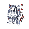 7p6rC 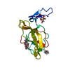 7p6sC 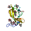 7p6tC  7pfpC C: citing same article ( M: map data used to model this data |
|---|---|
| Similar structure data |


-Links
 PDBj
PDBj


- Assembly
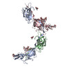
| Deposited unit | 
|
|---|---|
| 1 |
|
-Components
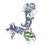
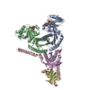
-Protein , 2 types, 2 molecules UF
| #1: Protein | Mass: 61498.816 Da / Num. of mol.: 1 / Source method: isolated from a natural source / Source: (natural) Homo sapiens (human) / Plasmid details: Urine / References: UniProt: P07911 |
|---|---|
| #2: Protein | Mass: 18669.688 Da / Num. of mol.: 1 / Mutation: A27V Source method: isolated from a genetically manipulated source Source: (gene. exp.) Gene: fimH, UTI89_C5017 / Plasmid: pD441-SR / Production host: |
-Sugars , 4 types, 4 molecules
| #3: Polysaccharide | 2-acetamido-2-deoxy-beta-D-glucopyranose-(1-4)-[alpha-L-fucopyranose-(1-6)]2-acetamido-2-deoxy-beta- ...2-acetamido-2-deoxy-beta-D-glucopyranose-(1-4)-[alpha-L-fucopyranose-(1-6)]2-acetamido-2-deoxy-beta-D-glucopyranose Source method: isolated from a genetically manipulated source |
|---|---|
| #4: Polysaccharide | 2-acetamido-2-deoxy-beta-D-glucopyranose-(1-4)-2-acetamido-2-deoxy-beta-D-glucopyranose Source method: isolated from a genetically manipulated source |
| #5: Polysaccharide | beta-D-galactopyranose-(1-4)-2-acetamido-2-deoxy-beta-D-glucopyranose-(1-2)-alpha-D-mannopyranose- ...beta-D-galactopyranose-(1-4)-2-acetamido-2-deoxy-beta-D-glucopyranose-(1-2)-alpha-D-mannopyranose-(1-3)-[2-acetamido-2-deoxy-beta-D-glucopyranose-(1-2)-alpha-D-mannopyranose-(1-6)]beta-D-mannopyranose-(1-4)-2-acetamido-2-deoxy-beta-D-glucopyranose-(1-4)-2-acetamido-2-deoxy-beta-D-glucopyranose Type: oligosaccharide / Mass: 1479.349 Da / Num. of mol.: 1 Source method: isolated from a genetically manipulated source |
| #6: Polysaccharide | alpha-D-mannopyranose-(1-3)-[alpha-D-mannopyranose-(1-6)]alpha-D-mannopyranose-(1-6)-[alpha-D- ...alpha-D-mannopyranose-(1-3)-[alpha-D-mannopyranose-(1-6)]alpha-D-mannopyranose-(1-6)-[alpha-D-mannopyranose-(1-3)]beta-D-mannopyranose-(1-4)-2-acetamido-2-deoxy-beta-D-glucopyranose-(1-4)-2-acetamido-2-deoxy-beta-D-glucopyranose Source method: isolated from a genetically manipulated source |
-Details
| Has ligand of interest | Y |
|---|---|
| Has protein modification | Y |
-Experimental details
-Experiment
| Experiment | Method: ELECTRON MICROSCOPY |
|---|---|
| EM experiment | Aggregation state: FILAMENT / 3D reconstruction method: single particle reconstruction |
- Sample preparation
| Component |
| ||||||||||||||||||||||||
|---|---|---|---|---|---|---|---|---|---|---|---|---|---|---|---|---|---|---|---|---|---|---|---|---|---|
| Molecular weight | Experimental value: NO | ||||||||||||||||||||||||
| Source (natural) |
| ||||||||||||||||||||||||
| Source (recombinant) | Organism: | ||||||||||||||||||||||||
| Buffer solution | pH: 7 | ||||||||||||||||||||||||
| Buffer component | Conc.: 10 mM / Name: Na-HEPES / Formula: C8H17N2NaO4S | ||||||||||||||||||||||||
| Specimen | Conc.: 1.8 mg/ml / Embedding applied: NO / Shadowing applied: NO / Staining applied: NO / Vitrification applied: YES | ||||||||||||||||||||||||
| Specimen support | Grid material: COPPER / Grid mesh size: 300 divisions/in. / Grid type: Quantifoil R2/2 | ||||||||||||||||||||||||
| Vitrification | Instrument: FEI VITROBOT MARK IV / Cryogen name: ETHANE / Chamber temperature: 294 K |
- Electron microscopy imaging
| Experimental equipment |  Model: Titan Krios / Image courtesy: FEI Company |
|---|---|
| Microscopy | Model: TFS KRIOS |
| Electron gun | Electron source:  FIELD EMISSION GUN / Accelerating voltage: 300 kV / Illumination mode: FLOOD BEAM FIELD EMISSION GUN / Accelerating voltage: 300 kV / Illumination mode: FLOOD BEAM |
| Electron lens | Mode: BRIGHT FIELD / Cs: 2.7 mm / Alignment procedure: COMA FREE |
| Specimen holder | Cryogen: NITROGEN / Specimen holder model: FEI TITAN KRIOS AUTOGRID HOLDER |
| Image recording | Average exposure time: 1.7 sec. / Electron dose: 40 e/Å2 / Film or detector model: GATAN K3 BIOQUANTUM (6k x 4k) / Num. of real images: 13616 |
| EM imaging optics | Energyfilter name: GIF Bioquantum / Energyfilter slit width: 20 eV |
- Processing
| Software |
| |||||||||||||||||||||||||||||||||||||||||||||
|---|---|---|---|---|---|---|---|---|---|---|---|---|---|---|---|---|---|---|---|---|---|---|---|---|---|---|---|---|---|---|---|---|---|---|---|---|---|---|---|---|---|---|---|---|---|---|
| EM software |
| |||||||||||||||||||||||||||||||||||||||||||||
| CTF correction | Type: PHASE FLIPPING AND AMPLITUDE CORRECTION | |||||||||||||||||||||||||||||||||||||||||||||
| Particle selection | Num. of particles selected: 3767790 | |||||||||||||||||||||||||||||||||||||||||||||
| 3D reconstruction | Resolution: 7.4 Å / Resolution method: FSC 0.143 CUT-OFF / Num. of particles: 225819 / Num. of class averages: 1 / Symmetry type: POINT | |||||||||||||||||||||||||||||||||||||||||||||
| Atomic model building | B value: 396.23 / Protocol: RIGID BODY FIT / Space: REAL | |||||||||||||||||||||||||||||||||||||||||||||
| Atomic model building | 3D fitting-ID: 1 / Source name: PDB / Type: experimental model
| |||||||||||||||||||||||||||||||||||||||||||||
| Refinement | Cross valid method: NONE Stereochemistry target values: GeoStd + Monomer Library + CDL v1.2 | |||||||||||||||||||||||||||||||||||||||||||||
| Displacement parameters | Biso mean: 396.23 Å2 | |||||||||||||||||||||||||||||||||||||||||||||
| Refine LS restraints |
|
