 Movie
Movie Controller


+ Open data

- Basic information
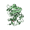
| Entry | Database: PDB / ID: 7pu1 | ||||||||||||
|---|---|---|---|---|---|---|---|---|---|---|---|---|---|
| Title | High resolution X-ray structure of Thermoascus aurantiacus LPMO | ||||||||||||
 Components Components | Gh61 isozyme a | ||||||||||||
 Keywords Keywords | OXIDOREDUCTASE / Copper binding protein / Beta-sandwich fold / Auxillary | ||||||||||||
| Function / homology |  Function and homology information Function and homology informationlytic cellulose monooxygenase (C4-dehydrogenating) / cellulase activity / cellulose catabolic process / monooxygenase activity / extracellular region / metal ion binding Similarity search - Function | ||||||||||||
| Biological species |  Thermoascus aurantiacus (fungus) Thermoascus aurantiacus (fungus) | ||||||||||||
| Method |  X-RAY DIFFRACTION / SYNCHROTRON / MOLECULAR REPLACEMENT / Resolution: 1.06 Å X-RAY DIFFRACTION / SYNCHROTRON / MOLECULAR REPLACEMENT / Resolution: 1.06 Å | ||||||||||||
 Authors Authors | Banerjee, S. / Frandsen, K.E.H. / Singh, R.K. / Bjerrum, M.J. / Lo Leggio, L. | ||||||||||||
| Funding support |  Sweden, 3items Sweden, 3items
| ||||||||||||
 Citation Citation | Journal: Biomolecules / Year: 2022 Title: Protonation State of an Important Histidine from High Resolution Structures of Lytic Polysaccharide Monooxygenases. Authors: Banerjee, S. / Muderspach, S.J. / Tandrup, T. / Frandsen, K.E.H. / Singh, R.K. / Ipsen, J.O. / Hernandez-Rollan, C. / Norholm, M.H.H. / Bjerrum, M.J. / Johansen, K.S. / Lo Leggio, L. | ||||||||||||
| History |
|
- Structure visualization
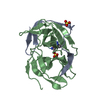
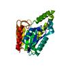
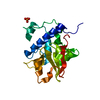
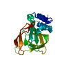
| Structure viewer | Molecule: MolmilJmol/JSmol |
|---|
- Downloads & links
-Download
| PDBx/mmCIF format | 7pu1.cif.gz | 207.5 KB | Display | PDBx/mmCIF format |
|---|---|---|---|---|
| PDB format | pdb7pu1.ent.gz | Display | PDB format | |
| PDBx/mmJSON format | 7pu1.json.gz | Tree view | PDBx/mmJSON format | |
| Others |  Other downloads Other downloads |
-Validation report
| Summary document | 7pu1_validation.pdf.gz | 466.2 KB | Display | wwPDB validaton report |
|---|---|---|---|---|
| Full document | 7pu1_full_validation.pdf.gz | 474.9 KB | Display | |
| Data in XML | 7pu1_validation.xml.gz | 28.7 KB | Display | |
| Data in CIF | 7pu1_validation.cif.gz | 43.9 KB | Display | |
| Arichive directory | https://data.pdbj.org/pub/pdb/validation_reports/pu/7pu1ftp://data.pdbj.org/pub/pdb/validation_reports/pu/7pu1 | HTTPS FTP |
-Related structure data
| Related structure data |  7ptzC  2yetS C: citing same article ( S: Starting model for refinement |
|---|---|
| Similar structure data |


-Links
 PDBj
PDBj- Assembly
| Deposited unit | 
| ||||||||
|---|---|---|---|---|---|---|---|---|---|
| 1 | 
| ||||||||
| 2 | 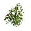
| ||||||||
| Unit cell |
| ||||||||
| Noncrystallographic symmetry (NCS) | NCS domain: (Details: Chains AAA BBB) |
-Components
| #1: Protein | Mass: 24418.043 Da / Num. of mol.: 2 Source method: isolated from a genetically manipulated source Source: (gene. exp.) Thermoascus aurantiacus (fungus) / Production host: #2: Chemical |   Mass: 63.546 Da / Num. of mol.: 2 / Source method: obtained synthetically / Formula: Cu Mass: 63.546 Da / Num. of mol.: 2 / Source method: obtained synthetically / Formula: Cu#3: Sugar |   Type: D-saccharide, beta linking / Mass: 221.208 Da / Num. of mol.: 2 / Source method: obtained synthetically / Formula: C8H15NO6 Type: D-saccharide, beta linking / Mass: 221.208 Da / Num. of mol.: 2 / Source method: obtained synthetically / Formula: C8H15NO6#4: Chemical | ChemComp-CL /   Mass: 35.453 Da / Num. of mol.: 19 / Source method: isolated from a natural source / Formula: Cl Mass: 35.453 Da / Num. of mol.: 19 / Source method: isolated from a natural source / Formula: Cl#5: Water | ChemComp-HOH / |  Mass: 18.015 Da / Num. of mol.: 705 / Source method: isolated from a natural source / Formula: H2O Mass: 18.015 Da / Num. of mol.: 705 / Source method: isolated from a natural source / Formula: H2OHas ligand of interest | N | |
|---|
-Experimental details
-Experiment
| Experiment | Method: X-RAY DIFFRACTION / Number of used crystals: 1 |
|---|
- Sample preparation
| Crystal | Density Matthews: 2.35 Å3/Da / Density % sol: 47.75 % |
|---|---|
| Crystal grow | Temperature: 293 K / Method: vapor diffusion, sitting drop / pH: 7.5 Details: 0.1 M HEPES pH 7.5, 20m M MgCl2 and 22 %(w/v) polyacrylic acid 5100 sodium salt |
-Data collection
| Diffraction | Mean temperature: 100 K / Serial crystal experiment: N |
|---|---|
| Diffraction source | Source: SYNCHROTRON / Site: MAX IV / Beamline: BioMAX / Wavelength: 0.9538 Å |
| Detector | Type: DECTRIS EIGER X 16M / Detector: PIXEL / Date: Jun 26, 2020 |
| Radiation | Protocol: SINGLE WAVELENGTH / Monochromatic (M) / Laue (L): M / Scattering type: x-ray |
| Radiation wavelength | Wavelength: 0.9538 Å / Relative weight: 1 |
| Reflection | Resolution: 1.06→37.34 Å / Num. obs: 171807 / % possible obs: 84.2 % / Redundancy: 6.6 % / CC1/2: 0.99 / Rrim(I) all: 0.054 / Net I/σ(I): 18.57 |
| Reflection shell | Resolution: 1.06→1.1 Å / Mean I/σ(I) obs: 3.6 / Num. unique obs: 12524 / CC1/2: 0.89 / Rrim(I) all: 0.44 |
- Processing
| Software |
| ||||||||||||||||||||||||||||||||||||||||||||||||||||||||||||||||||||||||||||||||||||||||||||||||||||||||||||||||||||||||||||||||||||||||||||||||||||||||||||||||
|---|---|---|---|---|---|---|---|---|---|---|---|---|---|---|---|---|---|---|---|---|---|---|---|---|---|---|---|---|---|---|---|---|---|---|---|---|---|---|---|---|---|---|---|---|---|---|---|---|---|---|---|---|---|---|---|---|---|---|---|---|---|---|---|---|---|---|---|---|---|---|---|---|---|---|---|---|---|---|---|---|---|---|---|---|---|---|---|---|---|---|---|---|---|---|---|---|---|---|---|---|---|---|---|---|---|---|---|---|---|---|---|---|---|---|---|---|---|---|---|---|---|---|---|---|---|---|---|---|---|---|---|---|---|---|---|---|---|---|---|---|---|---|---|---|---|---|---|---|---|---|---|---|---|---|---|---|---|---|---|---|---|
| Refinement | Method to determine structure: MOLECULAR REPLACEMENT Starting model: 2YET Resolution: 1.06→37.37 Å / Cor.coef. Fo:Fc: 0.983 / Cor.coef. Fo:Fc free: 0.979 / WRfactor Rfree: 0.144 / WRfactor Rwork: 0.118 / SU B: 0.651 / SU ML: 0.014 / Average fsc free: 0.9715 / Average fsc work: 0.9751 / Cross valid method: FREE R-VALUE / ESU R: 0.025 / ESU R Free: 0.026 Details: Hydrogens have been added in their riding positions
| ||||||||||||||||||||||||||||||||||||||||||||||||||||||||||||||||||||||||||||||||||||||||||||||||||||||||||||||||||||||||||||||||||||||||||||||||||||||||||||||||
| Solvent computation | Ion probe radii: 0.8 Å / Shrinkage radii: 0.8 Å / VDW probe radii: 1.2 Å / Solvent model: MASK BULK SOLVENT | ||||||||||||||||||||||||||||||||||||||||||||||||||||||||||||||||||||||||||||||||||||||||||||||||||||||||||||||||||||||||||||||||||||||||||||||||||||||||||||||||
| Displacement parameters | Biso mean: 12.983 Å2
| ||||||||||||||||||||||||||||||||||||||||||||||||||||||||||||||||||||||||||||||||||||||||||||||||||||||||||||||||||||||||||||||||||||||||||||||||||||||||||||||||
| Refinement step | Cycle: LAST / Resolution: 1.06→37.37 Å
| ||||||||||||||||||||||||||||||||||||||||||||||||||||||||||||||||||||||||||||||||||||||||||||||||||||||||||||||||||||||||||||||||||||||||||||||||||||||||||||||||
| Refine LS restraints |
| ||||||||||||||||||||||||||||||||||||||||||||||||||||||||||||||||||||||||||||||||||||||||||||||||||||||||||||||||||||||||||||||||||||||||||||||||||||||||||||||||
| LS refinement shell |
|
