[English] 日本語
Yorodumi
- PDB-2q30: Crystal structure of a rmlc-like cupin protein (dde_2303) from de... -

+
Open data


ID or keywords:

Loading...

-
Basic information

Entry
Database: PDB / ID: 2q30
TitleCrystal structure of a rmlc-like cupin protein (dde_2303) from desulfovibrio desulfuricans subsp. at 1.94 A resolution
ComponentsUncharacterized protein
KeywordsUNKNOWN FUNCTION / Double-stranded beta-helix fold / structural genomics / Joint Center for Structural Genomics / JCSG / Protein Structure Initiative / PSI-2
Function / homologyCupin 2, conserved barrel / Cupin domain / RmlC-like cupin domain superfamily / Jelly Rolls / RmlC-like jelly roll fold / Jelly Rolls / Sandwich / Mainly Beta / Cupin 2 conserved barrel domain protein
Function and homology information
Biological speciesDesulfovibrio desulfuricans subsp. desulfuricans str. G20 (bacteria)
MethodX-RAY DIFFRACTION / SYNCHROTRON / SAD / Resolution: 1.94 Å
AuthorsJoint Center for Structural Genomics (JCSG)
CitationJournal: To be published
Title: Crystal structure of uncharacterized protein (YP_388795.1) from Desulfovibrio desulfuricans G20 at 1.94 A resolution
Authors: Joint Center for Structural Genomics (JCSG)
History
DepositionMay 29, 2007Deposition site: RCSB / Processing site: RCSB
Revision 1.0Jun 19, 2007Provider: repository / Type: Initial release
Revision 1.1May 1, 2008Group: Version format compliance
Revision 1.2Jul 13, 2011Group: Advisory / Version format compliance
Revision 1.3Oct 18, 2017Group: Refinement description / Category: software / Item: _software.classification / _software.name
Revision 1.4Jul 24, 2019Group: Data collection / Derived calculations / Refinement description
Category: software / struct_conn
Item: _software.classification / _software.contact_author ..._software.classification / _software.contact_author / _software.contact_author_email / _software.language / _software.location / _software.name / _software.type / _software.version / _struct_conn.pdbx_leaving_atom_flag
Revision 1.5Jan 25, 2023Group: Database references / Derived calculations / Category: database_2 / struct_ref_seq_dif / struct_site
Item: _database_2.pdbx_DOI / _database_2.pdbx_database_accession ..._database_2.pdbx_DOI / _database_2.pdbx_database_accession / _struct_ref_seq_dif.details / _struct_site.pdbx_auth_asym_id / _struct_site.pdbx_auth_comp_id / _struct_site.pdbx_auth_seq_id
Revision 1.6Nov 13, 2024Group: Data collection / Refinement description / Structure summary
Category: chem_comp_atom / chem_comp_bond ...chem_comp_atom / chem_comp_bond / pdbx_entry_details / pdbx_modification_feature / struct_ncs_dom_lim
Item: _struct_ncs_dom_lim.beg_auth_comp_id / _struct_ncs_dom_lim.end_auth_comp_id
Remark 300 
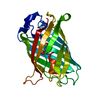
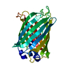
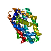
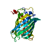
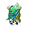
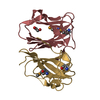
BIOMOLECULE: 1,2,3,4 THIS ENTRY CONTAINS THE CRYSTALLOGRAPHIC ASYMMETRIC UNIT WHICH CONSISTS OF 8 ... BIOMOLECULE: 1,2,3,4 THIS ENTRY CONTAINS THE CRYSTALLOGRAPHIC ASYMMETRIC UNIT WHICH CONSISTS OF 8 CHAINS. SEE REMARK 350 FOR INFORMATION ON GENERATING THE BIOLOGICAL MOLECULE(S). EBI/PISA ANALYSIS SUPPORTS THE ASSIGNMENT OF A DIMER AS THE SIGNIFICANT OLIGOMERIZATION STATE.
Remark 999 SEQUENCE THE CONSTRUCT WAS EXPRESSED WITH A PURIFICATION TAG MGSDKIHHHHHHENLYFQG. THE TAG WAS ... SEQUENCE THE CONSTRUCT WAS EXPRESSED WITH A PURIFICATION TAG MGSDKIHHHHHHENLYFQG. THE TAG WAS REMOVED WITH TEV PROTEASE LEAVING ONLY A GLYCINE (0) FOLLOWED BY THE TARGET SEQUENCE.

-
Structure visualization

Structure viewerMolecule:
MolmilJmol/JSmol

Downloads & links

-
Assembly

Deposited unit
A: Uncharacterized protein
B: Uncharacterized protein
C: Uncharacterized protein
D: Uncharacterized protein
E: Uncharacterized protein
F: Uncharacterized protein
G: Uncharacterized protein
H: Uncharacterized protein
hetero molecules


Theoretical massNumber of molelcules
Total (without water)97,75716
Polymers97,2268
Non-polymers5318
Water11,782654
1
A: Uncharacterized protein
B: Uncharacterized protein
hetero molecules


Theoretical massNumber of molelcules
Total (without water)24,4314
Polymers24,3072
Non-polymers1242
Water362
TypeNameSymmetry operationNumber
identity operation1_555x,y,z1
Buried area3240 Å2
ΔGint-15 kcal/mol
Surface area9410 Å2
MethodPISA
2
C: Uncharacterized protein
D: Uncharacterized protein
hetero molecules


Theoretical massNumber of molelcules
Total (without water)24,5275
Polymers24,3072
Non-polymers2203
Water362
TypeNameSymmetry operationNumber
identity operation1_555x,y,z1
Buried area3490 Å2
ΔGint-34 kcal/mol
Surface area9590 Å2
MethodPISA
3
E: Uncharacterized protein
F: Uncharacterized protein
hetero molecules


Theoretical massNumber of molelcules
Total (without water)24,4314
Polymers24,3072
Non-polymers1242
Water362
TypeNameSymmetry operationNumber
identity operation1_555x,y,z1
Buried area3080 Å2
ΔGint-6 kcal/mol
Surface area9720 Å2
MethodPISA
4
G: Uncharacterized protein
H: Uncharacterized protein
hetero molecules


Theoretical massNumber of molelcules
Total (without water)24,3693
Polymers24,3072
Non-polymers621
Water362
TypeNameSymmetry operationNumber
identity operation1_555x,y,z1
Buried area2790 Å2
ΔGint-9 kcal/mol
Surface area9590 Å2
MethodPISA
Unit cell
Length a, b, c (Å)43.140, 133.950, 74.660
Angle α, β, γ (deg.)90.000, 93.040, 90.000
Int Tables number4
Space group name H-MP1211
Noncrystallographic symmetry (NCS)NCS domain:
IDEns-IDDetails
11A
21B
31C
41D
51E
61F
71G
81H

NCS domain segments:

Component-ID: 1 / Ens-ID: 1 / Beg auth comp-ID: LYS / Beg label comp-ID: LYS / End auth comp-ID: PRO / End label comp-ID: PRO / Refine code: 4 / Auth seq-ID: 6 - 108 / Label seq-ID: 7 - 109

Dom-IDAuth asym-IDLabel asym-ID
1AA
2BB
3CC
4DD
5EE
6FF
7GG
8HH

-
Components

#1: Protein
Uncharacterized protein


Mass: 12153.260 Da / Num. of mol.: 8
Source method: isolated from a genetically manipulated source
Source: (gene. exp.) Desulfovibrio desulfuricans subsp. desulfuricans str. G20 (bacteria)
Species: Desulfovibrio desulfuricans / Strain: subsp. desulfuricans str. G20 / Gene: YP_388795.1, Dde_2303 / Plasmid: speedET / Production host: Escherichia coli (E. coli) / Strain (production host): HK100 / References: UniProt: Q30YZ6
#2: Chemical
ChemComp-EDO / 1,2-ETHANEDIOL / ETHYLENE GLYCOL


Mass: 62.068 Da / Num. of mol.: 7 / Source method: obtained synthetically / Formula: C2H6O2
#3: Chemical ChemComp-SO4 / SULFATE ION


Mass: 96.063 Da / Num. of mol.: 1 / Source method: obtained synthetically / Formula: SO4
#4: Water ChemComp-HOH / water


Mass: 18.015 Da / Num. of mol.: 654 / Source method: isolated from a natural source / Formula: H2O
Has protein modificationY

-
Experimental details

-
Experiment

ExperimentMethod: X-RAY DIFFRACTION / Number of used crystals: 2

-
Sample preparation

Crystal
IDDensity Matthews3/Da)Density % sol (%)
12.2144.45
2
Crystal grow
Temperature (K)Crystal-IDMethodpHDetails
2931vapor diffusion, sitting drop7.5NANODROP, 35.0% PEG 3000, 0.2M Sodium chloride, 0.1M Tris-HCl pH 7.5, VAPOR DIFFUSION, SITTING DROP, temperature 293K
2932vapor diffusion, sitting drop8NANODROP, 40.0% PEG 3000, 0.2M Sodium chloride, 0.1M Tris-HCl pH 8.0, VAPOR DIFFUSION, SITTING DROP, temperature 293K

-
Data collection

Diffraction
IDMean temperature (K)Crystal-ID
11001
21002
Diffraction source
SourceSiteBeamlineIDWavelength (Å)
SYNCHROTRONALS 5.0.311
SYNCHROTRONALS 5.0.220.9803
Detector
TypeIDDetectorDateDetails
ADSC QUANTUM 3151CCDSep 10, 2006
ADSC QUANTUM 3152CCDSep 10, 2006Vertically collimating premirror, toroidal focusing hexapod mirror
Radiation
IDMonochromatorProtocolMonochromatic (M) / Laue (L)Scattering typeWavelength-ID
1Single crystal, cylindrically bent, asymmetrically cut Si(220) crystalSINGLE WAVELENGTHMx-ray1
2LN2 cooled double-crystal silicon (111)SINGLE WAVELENGTHMx-ray1
Radiation wavelength
IDWavelength (Å)Relative weight
111
20.98031
ReflectionResolution: 1.94→29.025 Å / Num. obs: 60310 / % possible obs: 81.5 % / Observed criterion σ(I): -3 / Biso Wilson estimate: 29.144 Å2 / Rmerge(I) obs: 0.06 / Net I/σ(I): 8.45
Reflection shell
Resolution (Å)Rmerge(I) obsMean I/σ(I) obsNum. measured obsNum. unique obsDiffraction-ID% possible all
1.94-2.010.3172.51180072011,257
2.01-2.090.2363.519617100641,281.5
2.09-2.180.194.21999298461,282.2
2.18-2.30.155.222535108101,282.8
2.3-2.440.1335.821975101761,283.5
2.44-2.630.1027.423637106381,284.2
2.63-2.90.0789.223742108451,284.6
2.9-3.310.05611.822591104761,285.6
3.31-4.170.0415.223160108731,286.4
4.17-29.0250.03216.722884109831,287.1

-
Phasing

PhasingMethod: SAD

-
Processing

Software
NameVersionClassificationNB
REFMAC5.2.0019refinement
PHENIXrefinement
SHELXphasing
MolProbity3beta29model building
XSCALEdata scaling
PDB_EXTRACT3data extraction
ADSCQuantumdata collection
XDSdata reduction
SHARPphasing
RefinementMethod to determine structure: SAD / Resolution: 1.94→29.025 Å / Cor.coef. Fo:Fc: 0.962 / Cor.coef. Fo:Fc free: 0.938 / SU B: 7.234 / SU ML: 0.106 / TLS residual ADP flag: LIKELY RESIDUAL / Cross valid method: THROUGHOUT / σ(F): 0 / ESU R: 0.166 / ESU R Free: 0.15
Stereochemistry target values: MAXIMUM LIKELIHOOD WITH PHASES
Details: 1. HYDROGENS HAVE BEEN ADDED IN THE RIDING POSITIONS. 2. A MET-INHIBITION PROTOCOL WAS USED FOR SELENOMETHIONINE INCORPORATION DURING PROTEIN EXPRESSION. THE OCCUPANCY OF THE SE ATOMS IN THE ...Details: 1. HYDROGENS HAVE BEEN ADDED IN THE RIDING POSITIONS. 2. A MET-INHIBITION PROTOCOL WAS USED FOR SELENOMETHIONINE INCORPORATION DURING PROTEIN EXPRESSION. THE OCCUPANCY OF THE SE ATOMS IN THE MSE RESIDUES WAS REDUCED TO 0.75 FOR THE REDUCED SCATTERING POWER DUE TO PARTIAL S-MET INCORPORATION. 3. RESIDUES 1-3 IN ALL CHAINS, RESIDUE 4 IN CHAINS B AND C, AND RESIDUES 4-5 IN CHAINS E, F, G, AND H ARE DISORDERED AND NOT INCLUDED IN THE MODEL. 4. EDO AND SO4 MOLECULES FROM THE CRYSTALLIZATION/CRYO SOLUTION ARE MODELED. 5. ATOM RECORDS CONTAIN RESIDUAL B FACTORS ONLY.
RfactorNum. reflection% reflectionSelection details
Rfree0.212 3173 5.3 %RANDOM
Rwork0.167 ---
obs0.169 60283 96.41 %-
Solvent computationIon probe radii: 0.8 Å / Shrinkage radii: 0.8 Å / VDW probe radii: 1.2 Å / Solvent model: BABINET MODEL WITH MASK
Displacement parametersBiso mean: 26.755 Å2
Baniso -1Baniso -2Baniso -3
1-0.16 Å20 Å20.8 Å2
2---2.05 Å20 Å2
3---1.97 Å2
Refinement stepCycle: LAST / Resolution: 1.94→29.025 Å
ProteinNucleic acidLigandSolventTotal
Num. atoms6366 0 33 654 7053
Refine LS restraints
Refine-IDTypeDev idealDev ideal targetNumber
X-RAY DIFFRACTIONr_bond_refined_d0.0140.0226608
X-RAY DIFFRACTIONr_bond_other_d0.0050.024350
X-RAY DIFFRACTIONr_angle_refined_deg1.7911.9669005
X-RAY DIFFRACTIONr_angle_other_deg1.288310728
X-RAY DIFFRACTIONr_dihedral_angle_1_deg4.5615858
X-RAY DIFFRACTIONr_dihedral_angle_2_deg35.76724.712278
X-RAY DIFFRACTIONr_dihedral_angle_3_deg10.34151071
X-RAY DIFFRACTIONr_dihedral_angle_4_deg17.6091529
X-RAY DIFFRACTIONr_chiral_restr0.1050.21065
X-RAY DIFFRACTIONr_gen_planes_refined0.0060.027363
X-RAY DIFFRACTIONr_gen_planes_other0.0020.021226
X-RAY DIFFRACTIONr_nbd_refined0.160.21052
X-RAY DIFFRACTIONr_nbd_other0.1490.24497
X-RAY DIFFRACTIONr_nbtor_refined0.1530.23182
X-RAY DIFFRACTIONr_nbtor_other0.0770.23706
X-RAY DIFFRACTIONr_xyhbond_nbd_refined0.1050.2532
X-RAY DIFFRACTIONr_symmetry_vdw_refined0.1080.225
X-RAY DIFFRACTIONr_symmetry_vdw_other0.1510.271
X-RAY DIFFRACTIONr_symmetry_hbond_refined0.1160.222
X-RAY DIFFRACTIONr_mcbond_it1.95134695
X-RAY DIFFRACTIONr_mcbond_other0.58431689
X-RAY DIFFRACTIONr_mcangle_it2.30656893
X-RAY DIFFRACTIONr_scbond_it4.72482493
X-RAY DIFFRACTIONr_scangle_it6.636112098
Refine LS restraints NCS

Ens-ID: 1 / Number: 1175 / Refine-ID: X-RAY DIFFRACTION

Dom-IDAuth asym-IDTypeRms dev position (Å)Weight position
1AMEDIUM POSITIONAL0.30.5
2BMEDIUM POSITIONAL0.30.5
3CMEDIUM POSITIONAL0.220.5
4DMEDIUM POSITIONAL0.240.5
5EMEDIUM POSITIONAL0.230.5
6FMEDIUM POSITIONAL0.30.5
7GMEDIUM POSITIONAL0.450.5
8HMEDIUM POSITIONAL0.440.5
1AMEDIUM THERMAL0.942
2BMEDIUM THERMAL0.822
3CMEDIUM THERMAL0.892
4DMEDIUM THERMAL0.872
5EMEDIUM THERMAL0.932
6FMEDIUM THERMAL0.742
7GMEDIUM THERMAL0.822
8HMEDIUM THERMAL0.782
LS refinement shellResolution: 1.94→1.99 Å / Total num. of bins used: 20
RfactorNum. reflection% reflection
Rwork0.206 3476 -
obs--75.38 %
Refinement TLS params.

Method: refined / Refine-ID: X-RAY DIFFRACTION

IDL112)L122)L132)L222)L232)L332)S11 (Å °)S12 (Å °)S13 (Å °)S21 (Å °)S22 (Å °)S23 (Å °)S31 (Å °)S32 (Å °)S33 (Å °)T112)T122)T132)T222)T232)T332)Origin x (Å)Origin y (Å)Origin z (Å)
11.57380.4372-0.05862.32780.14720.687-0.0089-0.1054-0.08610.2762-0.01390.26360.0548-0.13180.0228-0.11250.00610.0281-0.04390.0282-0.12049.39810.8219.875
21.02370.21390.18641.9135-0.49211.23640.0002-0.00710.05740.10170.0056-0.0543-0.10560.0228-0.0058-0.14090.01230.0009-0.06240.0151-0.15121.74824.655.663
32.46620.69750.01641.8218-0.02420.59750.0527-0.0088-0.17920.1912-0.03090.12520.1353-0.0805-0.0218-0.09020.00710.0159-0.0419-0.0004-0.138727.8188.01144.707
41.42520.5066-0.10391.2073-0.03111.25060.06790.08250.19120.11240.00270.0294-0.12410.0048-0.0706-0.10840.00960.0092-0.03830.0185-0.124539.75222.49342.517
50.9048-0.60030.06393.6049-0.06471.1737-0.0852-0.06750.02610.06030.1227-0.106-0.0097-0.0492-0.0375-0.09280.00860.0232-0.0325-0.0076-0.077427.97561.3511.037
60.3493-0.6657-0.02974.3234-0.24511.4782-0.0636-0.147-0.13010.39080.23090.32950.0713-0.1724-0.1672-0.03450.0060.02310.01420.0365-0.034425.80943.45118.613
71.0811-0.1765-0.7493.4012-0.20192.65030.04570.05090.2022-0.14140.20990.1546-0.2293-0.1436-0.2557-0.07640.00330.0275-0.01160.05740.009246.4158.34351.974
80.669-0.2472-0.48393.2622-0.24871.7433-0.06010.01310.06030.09180.29250.4820.275-0.2424-0.2325-0.061-0.0397-0.0310.01310.0797-0.032643.48639.956.927
Refinement TLS group
IDRefine-IDRefine TLS-IDAuth asym-IDLabel asym-IDAuth seq-IDLabel seq-ID
1X-RAY DIFFRACTION1AA4 - 1095 - 110
2X-RAY DIFFRACTION2BB5 - 1096 - 110
3X-RAY DIFFRACTION3CC5 - 1096 - 110
4X-RAY DIFFRACTION4DD4 - 1095 - 110
5X-RAY DIFFRACTION5EE6 - 1097 - 110
6X-RAY DIFFRACTION6FF6 - 1097 - 110
7X-RAY DIFFRACTION7GG6 - 1097 - 110
8X-RAY DIFFRACTION8HH6 - 1097 - 110

+
About Yorodumi

-
News

-
Feb 9, 2022. New format data for meta-information of EMDB entries

New format data for meta-information of EMDB entries

  • Version 3 of the EMDB header file is now the official format.
  • The previous official version 1.9 will be removed from the archive.

Related info.:EMDB header

External links:wwPDB to switch to version 3 of the EMDB data model

-
Aug 12, 2020. Covid-19 info

Covid-19 info

URL: https://pdbj.org/emnavi/covid19.php

New page: Covid-19 featured information page in EM Navigator.

Related info.:Covid-19 info / Mar 5, 2020. Novel coronavirus structure data

+
Mar 5, 2020. Novel coronavirus structure data

Novel coronavirus structure data

Related info.:Yorodumi Speices / Aug 12, 2020. Covid-19 info

External links:COVID-19 featured content - PDBj / Molecule of the Month (242):Coronavirus Proteases

+
Jan 31, 2019. EMDB accession codes are about to change! (news from PDBe EMDB page)

EMDB accession codes are about to change! (news from PDBe EMDB page)

  • The allocation of 4 digits for EMDB accession codes will soon come to an end. Whilst these codes will remain in use, new EMDB accession codes will include an additional digit and will expand incrementally as the available range of codes is exhausted. The current 4-digit format prefixed with “EMD-” (i.e. EMD-XXXX) will advance to a 5-digit format (i.e. EMD-XXXXX), and so on. It is currently estimated that the 4-digit codes will be depleted around Spring 2019, at which point the 5-digit format will come into force.
  • The EM Navigator/Yorodumi systems omit the EMD- prefix.

Related info.:Q: What is EMD? / ID/Accession-code notation in Yorodumi/EM Navigator

External links:EMDB Accession Codes are Changing Soon! / Contact to PDBj

+
Jul 12, 2017. Major update of PDB

Major update of PDB

  • wwPDB released updated PDB data conforming to the new PDBx/mmCIF dictionary.
  • This is a major update changing the version number from 4 to 5, and with Remediation, in which all the entries are updated.
  • In this update, many items about electron microscopy experimental information are reorganized (e.g. em_software).
  • Now, EM Navigator and Yorodumi are based on the updated data.

External links:wwPDB Remediation / Enriched Model Files Conforming to OneDep Data Standards Now Available in the PDB FTP Archive

-
Yorodumi

Thousand views of thousand structures

  • Yorodumi is a browser for structure data from EMDB, PDB, SASBDB, etc.
  • This page is also the successor to EM Navigator detail page, and also detail information page/front-end page for Omokage search.
  • The word "yorodu" (or yorozu) is an old Japanese word meaning "ten thousand". "mi" (miru) is to see.

Related info.:EMDB / PDB / SASBDB / Comparison of 3 databanks / Yorodumi Search / Aug 31, 2016. New EM Navigator & Yorodumi / Yorodumi Papers / Jmol/JSmol / Function and homology information / Changes in new EM Navigator and Yorodumi

Read more