 Movie
Movie Controller

[English] 日本語
 Yorodumi
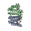
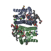
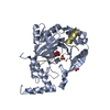
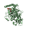
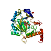
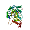
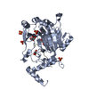
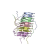
- PDB-7pow: Crystal structure of phosphatidyl serine synthase (PSS) in transi... -
+ Open data

- Basic information
| Entry | Database: PDB / ID: 7pow | ||||||
|---|---|---|---|---|---|---|---|
| Title | Crystal structure of phosphatidyl serine synthase (PSS) in transition state. | ||||||
 Components Components | CDP-diacylglycerol--serine O-phosphatidyltransferase | ||||||
 Keywords Keywords | LIPID BINDING PROTEIN / Membrane protein / Lipid synthase | ||||||
| Function / homology |  Function and homology information Function and homology informationCDP-diacylglycerol-serine O-phosphatidyltransferase / CDP-diacylglycerol-serine O-phosphatidyltransferase activity / phospholipid biosynthetic process / plasma membrane Similarity search - Function | ||||||
| Biological species |   Methanocaldococcus jannaschii (archaea) Methanocaldococcus jannaschii (archaea) | ||||||
| Method |  X-RAY DIFFRACTION / SYNCHROTRON / SAD / Resolution: 2.51 Å X-RAY DIFFRACTION / SYNCHROTRON / SAD / Resolution: 2.51 Å | ||||||
 Authors Authors | Yildiz, O. / Centola, M. | ||||||
| Funding support | 1items
| ||||||
 Citation Citation | Journal: Nat Commun / Year: 2021 Title: Crystal structures of phosphatidyl serine synthase PSS reveal the catalytic mechanism of CDP-DAG alcohol O-phosphatidyl transferases Authors: Centola, M. / Betz, H. / Yildiz, O. | ||||||
| History |
|
- Structure visualization
| Structure viewer | Molecule: MolmilJmol/JSmol |
|---|
- Downloads & links
-Download
| PDBx/mmCIF format | 7pow.cif.gz | 232.8 KB | Display | PDBx/mmCIF format |
|---|---|---|---|---|
| PDB format | pdb7pow.ent.gz | 154.2 KB | Display | PDB format |
| PDBx/mmJSON format | 7pow.json.gz | Tree view | PDBx/mmJSON format | |
| Others |  Other downloads Other downloads |
-Validation report
| Arichive directory | https://data.pdbj.org/pub/pdb/validation_reports/po/7powftp://data.pdbj.org/pub/pdb/validation_reports/po/7pow | HTTPS FTP |
|---|
-Related structure data


-Links
 PDBj
PDBj


- Assembly
| Deposited unit | 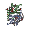
| ||||||||||||
|---|---|---|---|---|---|---|---|---|---|---|---|---|---|
| 1 |
| ||||||||||||
| Unit cell |
| ||||||||||||
| Components on special symmetry positions |
|
-Components
-Protein , 1 types, 2 molecules AB
| #1: Protein | Mass: 22520.367 Da / Num. of mol.: 2 Source method: isolated from a genetically manipulated source Source: (gene. exp.) Methanocaldococcus jannaschii (strain ATCC 43067 / DSM 2661 / JAL-1 / JCM 10045 / NBRC 100440) (archaea)Strain: ATCC 43067 / DSM 2661 / JAL-1 / JCM 10045 / NBRC 100440 Gene: pssA, MJ1212 / Production host:  References: UniProt: Q58609, CDP-diacylglycerol-serine O-phosphatidyltransferase |
|---|
-Non-polymers , 8 types, 97 molecules 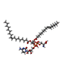


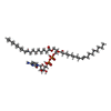


| #2: Chemical | ChemComp-SMW / ( Mass: 1111.240 Da / Num. of mol.: 1 / Source method: obtained synthetically / Formula: C51H92N4O18P2 / Feature type: SUBJECT OF INVESTIGATION Mass: 1111.240 Da / Num. of mol.: 1 / Source method: obtained synthetically / Formula: C51H92N4O18P2 / Feature type: SUBJECT OF INVESTIGATION | ||||||||||||
|---|---|---|---|---|---|---|---|---|---|---|---|---|---|
| #3: Chemical |  Mass: 40.078 Da / Num. of mol.: 2 / Source method: obtained synthetically / Formula: Ca Mass: 40.078 Da / Num. of mol.: 2 / Source method: obtained synthetically / Formula: Ca#4: Chemical |  Mass: 24.305 Da / Num. of mol.: 2 / Source method: obtained synthetically / Formula: Mg Mass: 24.305 Da / Num. of mol.: 2 / Source method: obtained synthetically / Formula: Mg#5: Chemical | ChemComp-CL /  Mass: 35.453 Da / Num. of mol.: 6 / Source method: obtained synthetically / Formula: Cl Mass: 35.453 Da / Num. of mol.: 6 / Source method: obtained synthetically / Formula: Cl#6: Chemical | ChemComp-OLC / (  Mass: 356.540 Da / Num. of mol.: 10 / Source method: obtained synthetically / Formula: C21H40O4 Mass: 356.540 Da / Num. of mol.: 10 / Source method: obtained synthetically / Formula: C21H40O4#7: Chemical | ChemComp-58A / |  Mass: 1006.147 Da / Num. of mol.: 1 / Source method: obtained synthetically / Formula: C48H85N3O15P2 / Feature type: SUBJECT OF INVESTIGATION Mass: 1006.147 Da / Num. of mol.: 1 / Source method: obtained synthetically / Formula: C48H85N3O15P2 / Feature type: SUBJECT OF INVESTIGATION#8: Chemical | ChemComp-SER / |  Type: L-peptide linking / Mass: 105.093 Da / Num. of mol.: 1 / Source method: obtained synthetically / Formula: C3H7NO3 Type: L-peptide linking / Mass: 105.093 Da / Num. of mol.: 1 / Source method: obtained synthetically / Formula: C3H7NO3#9: Water | ChemComp-HOH / | Mass: 18.015 Da / Num. of mol.: 74 / Source method: isolated from a natural source / Formula: H2O |
-Details
| Has ligand of interest | Y |
|---|---|
| Has protein modification | Y |
-Experimental details
-Experiment
| Experiment | Method: X-RAY DIFFRACTION / Number of used crystals: 1 |
|---|
- Sample preparation
| Crystal | Density Matthews: 2.55 Å3/Da / Density % sol: 51.85 % |
|---|---|
| Crystal grow | Temperature: 300 K / Method: lipidic cubic phase Details: Qiagens MBclass Suit and MBclass Suit II Molecular Dimension MemGold |
-Data collection
| Diffraction | Mean temperature: 100 K / Serial crystal experiment: N |
|---|---|
| Diffraction source | Source: SYNCHROTRON / Site: PETRA III, DESY  / Beamline: P11 / Wavelength: 0.9796 Å / Beamline: P11 / Wavelength: 0.9796 Å |
| Detector | Type: DECTRIS PILATUS 6M-F / Detector: PIXEL / Date: Aug 2, 2016 |
| Radiation | Protocol: SINGLE WAVELENGTH / Monochromatic (M) / Laue (L): M / Scattering type: x-ray |
| Radiation wavelength | Wavelength: 0.9796 Å / Relative weight: 1 |
| Reflection | Resolution: 2.5→38.04 Å / Num. obs: 30244 / % possible obs: 98.4 % / Redundancy: 5.4 % / Biso Wilson estimate: 24.95 Å2 / CC1/2: 0.987 / CC star: 0.997 / Rmerge(I) obs: 0.15 / Rpim(I) all: 0.05 / Rrim(I) all: 0.16 / Net I/σ(I): 9.2 |
| Reflection shell | Resolution: 2.5→2.6 Å / Redundancy: 3.44 % / Rmerge(I) obs: 0.29 / Mean I/σ(I) obs: 4.2 / Num. unique obs: 3027 / CC1/2: 0.966 / CC star: 0.991 / Rpim(I) all: 0.12 / Rrim(I) all: 0.31 / % possible all: 88.9 |
- Processing
| Software |
| ||||||||||||||||||||||||||||||||||||||||||||||||||||||||||||||||||||||||||||||||||||
|---|---|---|---|---|---|---|---|---|---|---|---|---|---|---|---|---|---|---|---|---|---|---|---|---|---|---|---|---|---|---|---|---|---|---|---|---|---|---|---|---|---|---|---|---|---|---|---|---|---|---|---|---|---|---|---|---|---|---|---|---|---|---|---|---|---|---|---|---|---|---|---|---|---|---|---|---|---|---|---|---|---|---|---|---|---|
| Refinement | Method to determine structure: SAD / Resolution: 2.51→38.04 Å / SU ML: 0.2592 / Cross valid method: FREE R-VALUE / σ(F): 1.27 / Phase error: 27.1958 Stereochemistry target values: GeoStd + Monomer Library + CDL v1.2
| ||||||||||||||||||||||||||||||||||||||||||||||||||||||||||||||||||||||||||||||||||||
| Solvent computation | Shrinkage radii: 0.9 Å / VDW probe radii: 1.11 Å / Solvent model: FLAT BULK SOLVENT MODEL | ||||||||||||||||||||||||||||||||||||||||||||||||||||||||||||||||||||||||||||||||||||
| Displacement parameters | Biso mean: 31.94 Å2 | ||||||||||||||||||||||||||||||||||||||||||||||||||||||||||||||||||||||||||||||||||||
| Refinement step | Cycle: LAST / Resolution: 2.51→38.04 Å
| ||||||||||||||||||||||||||||||||||||||||||||||||||||||||||||||||||||||||||||||||||||
| Refine LS restraints |
| ||||||||||||||||||||||||||||||||||||||||||||||||||||||||||||||||||||||||||||||||||||
| LS refinement shell |
| ||||||||||||||||||||||||||||||||||||||||||||||||||||||||||||||||||||||||||||||||||||
| Refinement TLS params. | Method: refined / Origin x: -5.922 Å / Origin y: 6.707 Å / Origin z: 49.666 Å
| ||||||||||||||||||||||||||||||||||||||||||||||||||||||||||||||||||||||||||||||||||||
| Refinement TLS group |
|
