 Movie
Movie Controller


+ Open data

- Basic information
| Entry | Database: PDB / ID: 7p9m | |||||||||
|---|---|---|---|---|---|---|---|---|---|---|
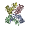
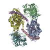
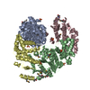
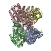
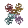
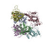
| Title | BrxU, GmrSD-family Type IV restriction enzyme | |||||||||
 Components Components | DUF262 domain-containing protein | |||||||||
 Keywords Keywords | DNA BINDING PROTEIN / Restriction endonuclease / Phage defence protein | |||||||||
| Function / homology | Domain of unknown function DUF262 / GmrSD restriction endonuclease, N-terminal domain / GmrSD restriction endonucleases N-terminal domain-containing protein Function and homology information Function and homology information | |||||||||
| Biological species |  | |||||||||
| Method |  X-RAY DIFFRACTION / SYNCHROTRON / SAD / Resolution: 2.85 Å X-RAY DIFFRACTION / SYNCHROTRON / SAD / Resolution: 2.85 Å | |||||||||
 Authors Authors | Picton, D.M. / Luyten, Y. / Morgan, R.D. / Nelson, A. / Smith, D.L. / Dryden, D.T.F. / Hinton, J.C.D. / Blower, T.R. | |||||||||
| Funding support |  United Kingdom, 2items United Kingdom, 2items
| |||||||||
 Citation Citation | Journal: Nucleic Acids Res. / Year: 2021 Title: The phage defence island of a multidrug resistant plasmid uses both BREX and type IV restriction for complementary protection from viruses. Authors: Picton, D.M. / Luyten, Y.A. / Morgan, R.D. / Nelson, A. / Smith, D.L. / Dryden, D.T.F. / Hinton, J.C.D. / Blower, T.R. | |||||||||
| History |
|
- Structure visualization
| Structure viewer | Molecule: MolmilJmol/JSmol |
|---|
- Downloads & links
-Download
| PDBx/mmCIF format | 7p9m.cif.gz | 252.1 KB | Display | PDBx/mmCIF format |
|---|---|---|---|---|
| PDB format | pdb7p9m.ent.gz | 186.8 KB | Display | PDB format |
| PDBx/mmJSON format | 7p9m.json.gz | Tree view | PDBx/mmJSON format | |
| Others |  Other downloads Other downloads |
-Validation report
| Arichive directory | https://data.pdbj.org/pub/pdb/validation_reports/p9/7p9mftp://data.pdbj.org/pub/pdb/validation_reports/p9/7p9m | HTTPS FTP |
|---|
-Related structure data


-Links
 PDBj
PDBj- Assembly
| Deposited unit | 
| ||||||||||||
|---|---|---|---|---|---|---|---|---|---|---|---|---|---|
| 1 |
| ||||||||||||
| Unit cell |
|
-Components
| #1: Protein | Mass: 67988.812 Da / Num. of mol.: 2 Source method: isolated from a genetically manipulated source Details: Plasmid pEFER Source: (gene. exp.) Strain: ATCC 35469 / DSM 13698 / CCUG 18766 / IAM 14443 / JCM 21226 / LMG 7866 / NBRC 102419 / NCTC 12128 / CDC 0568-73 Gene: EFER_p0024 / Production host: #2: Chemical |   Mass: 96.063 Da / Num. of mol.: 3 / Source method: obtained synthetically / Formula: SO4 Mass: 96.063 Da / Num. of mol.: 3 / Source method: obtained synthetically / Formula: SO4#3: Chemical | ChemComp-CL / |   Mass: 35.453 Da / Num. of mol.: 1 / Source method: obtained synthetically / Formula: Cl Mass: 35.453 Da / Num. of mol.: 1 / Source method: obtained synthetically / Formula: ClHas ligand of interest | N | |
|---|
-Experimental details
-Experiment
| Experiment | Method: X-RAY DIFFRACTION / Number of used crystals: 1 |
|---|
- Sample preparation
| Crystal | Density Matthews: 3.2 Å3/Da / Density % sol: 61.53 % / Description: Crystals looked like irregular shards |
|---|---|
| Crystal grow | Temperature: 294 K / Method: vapor diffusion, sitting drop / pH: 7.5 Details: 0.1 M Tris-HCl (pH 7.5), 0.2 M ammonium sulphate, 20% w/v PEG3350 PH range: 7.4-7.6 |
-Data collection
| Diffraction | Mean temperature: 100 K / Serial crystal experiment: N |
|---|---|
| Diffraction source | Source: SYNCHROTRON / Site: Diamond / Beamline: I04 / Wavelength: 0.9795 Å |
| Detector | Type: DECTRIS PILATUS3 6M / Detector: PIXEL / Date: Nov 23, 2020 |
| Radiation | Protocol: SINGLE WAVELENGTH / Monochromatic (M) / Laue (L): M / Scattering type: x-ray |
| Radiation wavelength | Wavelength: 0.9795 Å / Relative weight: 1 |
| Reflection | Resolution: 2.85→98.14 Å / Num. obs: 40622 / % possible obs: 99.8 % / Redundancy: 1.9 % / Biso Wilson estimate: 98.18 Å2 / CC1/2: 0.999 / Rmerge(I) obs: 0.044 / Net I/σ(I): 9.8 |
| Reflection shell | Resolution: 2.85→2.96 Å / Redundancy: 2 % / Rmerge(I) obs: 1.021 / Num. unique obs: 4514 / CC1/2: 0.368 / % possible all: 98.5 |
- Processing
| Software |
| ||||||||||||||||||||||||||||||||||||||||||||||||||||||||||||||||||||||||||||||||||||||||||||||||||
|---|---|---|---|---|---|---|---|---|---|---|---|---|---|---|---|---|---|---|---|---|---|---|---|---|---|---|---|---|---|---|---|---|---|---|---|---|---|---|---|---|---|---|---|---|---|---|---|---|---|---|---|---|---|---|---|---|---|---|---|---|---|---|---|---|---|---|---|---|---|---|---|---|---|---|---|---|---|---|---|---|---|---|---|---|---|---|---|---|---|---|---|---|---|---|---|---|---|---|---|
| Refinement | Method to determine structure: SAD / Resolution: 2.85→98.14 Å / SU ML: 0.6402 / Cross valid method: FREE R-VALUE / σ(F): 1.33 / Phase error: 37.6959 Stereochemistry target values: GeoStd + Monomer Library + CDL v1.2
| ||||||||||||||||||||||||||||||||||||||||||||||||||||||||||||||||||||||||||||||||||||||||||||||||||
| Solvent computation | Shrinkage radii: 0.9 Å / VDW probe radii: 1.11 Å / Solvent model: FLAT BULK SOLVENT MODEL | ||||||||||||||||||||||||||||||||||||||||||||||||||||||||||||||||||||||||||||||||||||||||||||||||||
| Displacement parameters | Biso mean: 97.24 Å2 | ||||||||||||||||||||||||||||||||||||||||||||||||||||||||||||||||||||||||||||||||||||||||||||||||||
| Refinement step | Cycle: LAST / Resolution: 2.85→98.14 Å
| ||||||||||||||||||||||||||||||||||||||||||||||||||||||||||||||||||||||||||||||||||||||||||||||||||
| Refine LS restraints |
| ||||||||||||||||||||||||||||||||||||||||||||||||||||||||||||||||||||||||||||||||||||||||||||||||||
| LS refinement shell |
|
