[English] 日本語
Yorodumi
- PDB-4aib: Crystal Structure of Ornithine Decarboxylase from Entamoeba histo... -

+
Open data


ID or keywords:

Loading...

-
Basic information

Entry
Database: PDB / ID: 4aib
TitleCrystal Structure of Ornithine Decarboxylase from Entamoeba histolytica.
ComponentsORNITHINE DECARBOXYLASE
KeywordsLYASE
Function / homology
Function and homology information


putrescine biosynthetic process from ornithine / ornithine decarboxylase / ornithine decarboxylase activity / cytoplasm
Similarity search - Function
Ornithine decarboxylase / Orn/DAP/Arg decarboxylase 2, conserved site / Orn/DAP/Arg decarboxylase 2, pyridoxal-phosphate binding site / Orn/DAP/Arg decarboxylases family 2 pyridoxal-P attachment site. / Orn/DAP/Arg decarboxylases family 2 signature 2. / Orn/DAP/Arg decarboxylase 2, C-terminal / Pyridoxal-dependent decarboxylase, C-terminal sheet domain / Ornithine/DAP/Arg decarboxylase / Orn/DAP/Arg decarboxylase 2, N-terminal / Pyridoxal-dependent decarboxylase, pyridoxal binding domain ...Ornithine decarboxylase / Orn/DAP/Arg decarboxylase 2, conserved site / Orn/DAP/Arg decarboxylase 2, pyridoxal-phosphate binding site / Orn/DAP/Arg decarboxylases family 2 pyridoxal-P attachment site. / Orn/DAP/Arg decarboxylases family 2 signature 2. / Orn/DAP/Arg decarboxylase 2, C-terminal / Pyridoxal-dependent decarboxylase, C-terminal sheet domain / Ornithine/DAP/Arg decarboxylase / Orn/DAP/Arg decarboxylase 2, N-terminal / Pyridoxal-dependent decarboxylase, pyridoxal binding domain / Lyase, Ornithine Decarboxylase; Chain A, domain 1 / Lyase, Ornithine Decarboxylase; Chain A, domain 1 / Alanine racemase / Alanine racemase/group IV decarboxylase, C-terminal / PLP-binding barrel / TIM Barrel / Alpha-Beta Barrel / Beta Barrel / Mainly Beta / Alpha Beta
Similarity search - Domain/homology
Ornithine decarboxylase
Similarity search - Component
Biological speciesENTAMOEBA HISTOLYTICA (eukaryote)
MethodX-RAY DIFFRACTION / MOLECULAR REPLACEMENT / Resolution: 2.87 Å
AuthorsPreeti, P. / Kumar, P. / Tomar, S.
CitationJournal: Plos One / Year: 2013
Title: Structural Insight Into Dfmo Resistant Ornithine Decarboxylase from Entamoeba Histolytica: An Inkling to Adaptive Evolution.
Authors: Preeti, P. / Tapas, S. / Kumar, P. / Tomar, S.
History
DepositionFeb 9, 2012Deposition site: PDBE / Processing site: PDBE
Revision 1.0Jan 30, 2013Provider: repository / Type: Initial release
Revision 1.1Dec 20, 2023Group: Data collection / Database references ...Data collection / Database references / Other / Refinement description
Category: chem_comp_atom / chem_comp_bond ...chem_comp_atom / chem_comp_bond / database_2 / pdbx_database_status / pdbx_initial_refinement_model
Item: _database_2.pdbx_DOI / _database_2.pdbx_database_accession / _pdbx_database_status.status_code_sf
Remark 700 
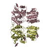
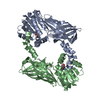
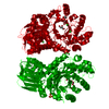
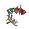
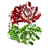
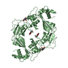
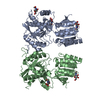
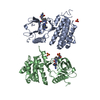
SHEET DETERMINATION METHOD: DSSP THE SHEETS PRESENTED AS "AC" IN EACH CHAIN ON SHEET RECORDS BELOW ... SHEET DETERMINATION METHOD: DSSP THE SHEETS PRESENTED AS "AC" IN EACH CHAIN ON SHEET RECORDS BELOW IS ACTUALLY AN 8-STRANDED BARREL THIS IS REPRESENTED BY A 9-STRANDED SHEET IN WHICH THE FIRST AND LAST STRANDS ARE IDENTICAL. THE SHEETS PRESENTED AS "BD" IN EACH CHAIN ON SHEET RECORDS BELOW IS ACTUALLY AN 8-STRANDED BARREL THIS IS REPRESENTED BY A 9-STRANDED SHEET IN WHICH THE FIRST AND LAST STRANDS ARE IDENTICAL. THE SHEETS PRESENTED AS "CB" IN EACH CHAIN ON SHEET RECORDS BELOW IS ACTUALLY AN 8-STRANDED BARREL THIS IS REPRESENTED BY A 9-STRANDED SHEET IN WHICH THE FIRST AND LAST STRANDS ARE IDENTICAL. THE SHEETS PRESENTED AS "DD" IN EACH CHAIN ON SHEET RECORDS BELOW IS ACTUALLY AN 8-STRANDED BARREL THIS IS REPRESENTED BY A 9-STRANDED SHEET IN WHICH THE FIRST AND LAST STRANDS ARE IDENTICAL.

-
Structure visualization

Structure viewerMolecule:
MolmilJmol/JSmol

Downloads & links

-
Assembly

Deposited unit
A: ORNITHINE DECARBOXYLASE
B: ORNITHINE DECARBOXYLASE
C: ORNITHINE DECARBOXYLASE
D: ORNITHINE DECARBOXYLASE


Theoretical massNumber of molelcules
Total (without water)178,2994
Polymers178,2994
Non-polymers00
Water1,820101
1
A: ORNITHINE DECARBOXYLASE
B: ORNITHINE DECARBOXYLASE


Theoretical massNumber of molelcules
Total (without water)89,1502
Polymers89,1502
Non-polymers00
Water362
TypeNameSymmetry operationNumber
identity operation1_555x,y,z1
Buried area3200 Å2
ΔGint-8.8 kcal/mol
Surface area30780 Å2
MethodPISA
2
C: ORNITHINE DECARBOXYLASE
D: ORNITHINE DECARBOXYLASE


Theoretical massNumber of molelcules
Total (without water)89,1502
Polymers89,1502
Non-polymers00
Water362
TypeNameSymmetry operationNumber
identity operation1_555x,y,z1
Buried area2290 Å2
ΔGint-6 kcal/mol
Surface area31610 Å2
MethodPISA
Unit cell
Length a, b, c (Å)76.671, 119.283, 179.292
Angle α, β, γ (deg.)90.00, 90.00, 90.00
Int Tables number19
Space group name H-MP212121

-
Components

#1: Protein
ORNITHINE DECARBOXYLASE /


Mass: 44574.781 Da / Num. of mol.: 4
Source method: isolated from a genetically manipulated source
Source: (gene. exp.) ENTAMOEBA HISTOLYTICA (eukaryote) / Strain: HM-1\:IMSS / Production host: ESCHERICHIA COLI (E. coli) / Strain (production host): BL21(DE3) / References: UniProt: Q58P26, ornithine decarboxylase
#2: Water ChemComp-HOH / water / Water


Mass: 18.015 Da / Num. of mol.: 101 / Source method: isolated from a natural source / Formula: H2O

-
Experimental details

-
Experiment

ExperimentMethod: X-RAY DIFFRACTION / Number of used crystals: 1

-
Sample preparation

CrystalDensity Matthews: 2.28 Å3/Da / Density % sol: 46.02 % / Description: NONE
Crystal growDetails: POLYETHYLENE GLYCOL, LITHIUM CHLORIDE

-
Data collection

DiffractionMean temperature: 100 K
Diffraction sourceSource: ROTATING ANODE / Type: BRUKER AXS MICROSTAR-H / Wavelength: 1.5418
DetectorType: MAR scanner 345 mm plate / Detector: IMAGE PLATE / Date: Jan 15, 2011 / Details: HELIOS
RadiationProtocol: SINGLE WAVELENGTH / Monochromatic (M) / Laue (L): M / Scattering type: x-ray
Radiation wavelengthWavelength: 1.5418 Å / Relative weight: 1
ReflectionResolution: 2.87→100 Å / Num. obs: 35570 / % possible obs: 92.1 % / Observed criterion σ(I): 2 / Redundancy: 3.4 % / Rmerge(I) obs: 0.15 / Net I/σ(I): 4.82
Reflection shellResolution: 2.87→2.92 Å / Redundancy: 2.1 % / Rmerge(I) obs: 0.54 / Mean I/σ(I) obs: 2 / % possible all: 59

-
Processing

Software
NameVersionClassification
REFMAC5.6.0117refinement
HKL-2000data reduction
SCALEPACKdata scaling
MOLREPphasing
RefinementMethod to determine structure: MOLECULAR REPLACEMENT
Starting model: PDB ENTRY 2ON3
Resolution: 2.87→99.5 Å / Cor.coef. Fo:Fc: 0.873 / Cor.coef. Fo:Fc free: 0.861 / SU B: 25.851 / SU ML: 0.451 / Cross valid method: THROUGHOUT / ESU R: 2.077 / ESU R Free: 0.504 / Stereochemistry target values: MAXIMUM LIKELIHOOD / Details: HYDROGENS HAVE BEEN ADDED IN THE RIDING POSITIONS.
RfactorNum. reflection% reflectionSelection details
Rfree0.29934 1763 5 %RANDOM
Rwork0.25266 ---
obs0.253 33646 91.79 %-
Solvent computationIon probe radii: 0.8 Å / Shrinkage radii: 0.8 Å / VDW probe radii: 1.2 Å / Solvent model: MASK
Displacement parametersBiso mean: 54.427 Å2
Baniso -1Baniso -2Baniso -3
1--1.23 Å20 Å20 Å2
2--1.21 Å20 Å2
3---0.03 Å2
Refinement stepCycle: LAST / Resolution: 2.87→99.5 Å
ProteinNucleic acidLigandSolventTotal
Num. atoms10484 0 0 101 10585
Refine LS restraints
Refine-IDTypeDev idealDev ideal targetNumber
X-RAY DIFFRACTIONr_bond_refined_d0.0050.0210701
X-RAY DIFFRACTIONr_bond_other_d
X-RAY DIFFRACTIONr_angle_refined_deg0.8311.96714423
X-RAY DIFFRACTIONr_angle_other_deg
X-RAY DIFFRACTIONr_dihedral_angle_1_deg3.76351289
X-RAY DIFFRACTIONr_dihedral_angle_2_deg36.19825.195487
X-RAY DIFFRACTIONr_dihedral_angle_3_deg14.866151954
X-RAY DIFFRACTIONr_dihedral_angle_4_deg7.8711536
X-RAY DIFFRACTIONr_chiral_restr0.0560.21596
X-RAY DIFFRACTIONr_gen_planes_refined0.0030.0217942
X-RAY DIFFRACTIONr_gen_planes_other
X-RAY DIFFRACTIONr_nbd_refined
X-RAY DIFFRACTIONr_nbd_other
X-RAY DIFFRACTIONr_nbtor_refined
X-RAY DIFFRACTIONr_nbtor_other
X-RAY DIFFRACTIONr_xyhbond_nbd_refined
X-RAY DIFFRACTIONr_xyhbond_nbd_other
X-RAY DIFFRACTIONr_metal_ion_refined
X-RAY DIFFRACTIONr_metal_ion_other
X-RAY DIFFRACTIONr_symmetry_vdw_refined
X-RAY DIFFRACTIONr_symmetry_vdw_other
X-RAY DIFFRACTIONr_symmetry_hbond_refined
X-RAY DIFFRACTIONr_symmetry_hbond_other
X-RAY DIFFRACTIONr_symmetry_metal_ion_refined
X-RAY DIFFRACTIONr_symmetry_metal_ion_other
X-RAY DIFFRACTIONr_mcbond_it
X-RAY DIFFRACTIONr_mcbond_other
X-RAY DIFFRACTIONr_mcangle_it
X-RAY DIFFRACTIONr_mcangle_other
X-RAY DIFFRACTIONr_scbond_it
X-RAY DIFFRACTIONr_scbond_other
X-RAY DIFFRACTIONr_scangle_it
X-RAY DIFFRACTIONr_scangle_other
X-RAY DIFFRACTIONr_long_range_B_refined
X-RAY DIFFRACTIONr_long_range_B_other
X-RAY DIFFRACTIONr_rigid_bond_restr
X-RAY DIFFRACTIONr_sphericity_free
X-RAY DIFFRACTIONr_sphericity_bonded
LS refinement shellResolution: 2.866→2.94 Å / Total num. of bins used: 20
RfactorNum. reflection% reflection
Rfree0.432 58 -
Rwork0.434 1540 -
obs--59.14 %

+
About Yorodumi

-
News

-
Feb 9, 2022. New format data for meta-information of EMDB entries

New format data for meta-information of EMDB entries

  • Version 3 of the EMDB header file is now the official format.
  • The previous official version 1.9 will be removed from the archive.

Related info.:EMDB header

External links:wwPDB to switch to version 3 of the EMDB data model

-
Aug 12, 2020. Covid-19 info

Covid-19 info

URL: https://pdbj.org/emnavi/covid19.php

New page: Covid-19 featured information page in EM Navigator.

Related info.:Covid-19 info / Mar 5, 2020. Novel coronavirus structure data

+
Mar 5, 2020. Novel coronavirus structure data

Novel coronavirus structure data

Related info.:Yorodumi Speices / Aug 12, 2020. Covid-19 info

External links:COVID-19 featured content - PDBj / Molecule of the Month (242):Coronavirus Proteases

+
Jan 31, 2019. EMDB accession codes are about to change! (news from PDBe EMDB page)

EMDB accession codes are about to change! (news from PDBe EMDB page)

  • The allocation of 4 digits for EMDB accession codes will soon come to an end. Whilst these codes will remain in use, new EMDB accession codes will include an additional digit and will expand incrementally as the available range of codes is exhausted. The current 4-digit format prefixed with “EMD-” (i.e. EMD-XXXX) will advance to a 5-digit format (i.e. EMD-XXXXX), and so on. It is currently estimated that the 4-digit codes will be depleted around Spring 2019, at which point the 5-digit format will come into force.
  • The EM Navigator/Yorodumi systems omit the EMD- prefix.

Related info.:Q: What is EMD? / ID/Accession-code notation in Yorodumi/EM Navigator

External links:EMDB Accession Codes are Changing Soon! / Contact to PDBj

+
Jul 12, 2017. Major update of PDB

Major update of PDB

  • wwPDB released updated PDB data conforming to the new PDBx/mmCIF dictionary.
  • This is a major update changing the version number from 4 to 5, and with Remediation, in which all the entries are updated.
  • In this update, many items about electron microscopy experimental information are reorganized (e.g. em_software).
  • Now, EM Navigator and Yorodumi are based on the updated data.

External links:wwPDB Remediation / Enriched Model Files Conforming to OneDep Data Standards Now Available in the PDB FTP Archive

-
Yorodumi

Thousand views of thousand structures

  • Yorodumi is a browser for structure data from EMDB, PDB, SASBDB, etc.
  • This page is also the successor to EM Navigator detail page, and also detail information page/front-end page for Omokage search.
  • The word "yorodu" (or yorozu) is an old Japanese word meaning "ten thousand". "mi" (miru) is to see.

Related info.:EMDB / PDB / SASBDB / Comparison of 3 databanks / Yorodumi Search / Aug 31, 2016. New EM Navigator & Yorodumi / Yorodumi Papers / Jmol/JSmol / Function and homology information / Changes in new EM Navigator and Yorodumi

Read more