 Movie
Movie Controller

[English] 日本語
 Yorodumi
- PDB-7p97: Structure of 3-phospho-D-glycerate guanylyltransferase with produ... -
+ Open data

- Basic information
| Entry | Database: PDB / ID: 7p97 | |||||||||
|---|---|---|---|---|---|---|---|---|---|---|
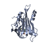
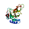
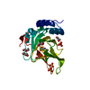
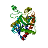
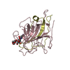
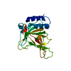
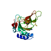
| Title | Structure of 3-phospho-D-glycerate guanylyltransferase with product 3-GPPG bound | |||||||||
 Components Components | 3-phospho-D-glycerate guanylyltransferase | |||||||||
 Keywords Keywords | TRANSFERASE / F420 synthesis / FbiD / CofC / guanylyltransferase / 3-phospho-glycerate | |||||||||
| Function / homology | 3-phospho-D-glycerate guanylyltransferase / phospholactate guanylyltransferase activity / Phosphoenolpyruvate guanylyltransferase CofC / Guanylyl transferase CofC like / F420-0 metabolic process / Nucleotide-diphospho-sugar transferases / GTP binding / 3-(guanosine-5'-diphospho)-D-glycerate / 3-phospho-D-glycerate guanylyltransferase Function and homology information Function and homology information | |||||||||
| Biological species |  Mycetohabitans rhizoxinica (bacteria) Mycetohabitans rhizoxinica (bacteria) | |||||||||
| Method |  X-RAY DIFFRACTION / SYNCHROTRON / MOLECULAR REPLACEMENT / Resolution: 2.35 Å X-RAY DIFFRACTION / SYNCHROTRON / MOLECULAR REPLACEMENT / Resolution: 2.35 Å | |||||||||
 Authors Authors | Palm, G.J. / Berndt, L. / Lammers, M. | |||||||||
| Funding support |  Germany, 2items Germany, 2items
| |||||||||
 Citation Citation | Journal: Mbio / Year: 2022 Title: Diversification by CofC and Control by CofD Govern Biosynthesis and Evolution of Coenzyme F 420 and Its Derivative 3PG-F 420. Authors: Hasan, M. / Schulze, S. / Berndt, L. / Palm, G.J. / Braga, D. / Richter, I. / Last, D. / Lammers, M. / Lackner, G. | |||||||||
| History |
|
- Structure visualization
| Structure viewer | Molecule: MolmilJmol/JSmol |
|---|
- Downloads & links
-Download
| PDBx/mmCIF format | 7p97.cif.gz | 292.7 KB | Display | PDBx/mmCIF format |
|---|---|---|---|---|
| PDB format | pdb7p97.ent.gz | Display | PDB format | |
| PDBx/mmJSON format | 7p97.json.gz | Tree view | PDBx/mmJSON format | |
| Others |  Other downloads Other downloads |
-Validation report
| Arichive directory | https://data.pdbj.org/pub/pdb/validation_reports/p9/7p97ftp://data.pdbj.org/pub/pdb/validation_reports/p9/7p97 | HTTPS FTP |
|---|
-Related structure data
| Related structure data | 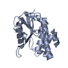 6bwgS S: Starting model for refinement |
|---|---|
| Similar structure data |


-Links
 PDBj
PDBj

- Assembly
| Deposited unit | 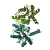
| |||||||||
|---|---|---|---|---|---|---|---|---|---|---|
| 1 | 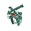
| |||||||||
| 2 | 
| |||||||||
| Unit cell |
| |||||||||
| Components on special symmetry positions |
|
-Components
| #1: Protein | Mass: 25906.516 Da / Num. of mol.: 2 Source method: isolated from a genetically manipulated source Source: (gene. exp.) Mycetohabitans rhizoxinica (strain DSM 19002 / CIP 109453 / HKI 454) (bacteria)Strain: DSM 19002 / CIP 109453 / HKI 454 / Gene: cofC, RBRH_02550 / Plasmid: pACYCduet / Details (production host): N-terminal His-tag / Production host: References: UniProt: E5ASS2, 3-phospho-D-glycerate guanylyltransferase #2: Chemical |   Mass: 531.263 Da / Num. of mol.: 2 / Source method: obtained synthetically / Formula: C13H19N5O14P2 / Feature type: SUBJECT OF INVESTIGATION Mass: 531.263 Da / Num. of mol.: 2 / Source method: obtained synthetically / Formula: C13H19N5O14P2 / Feature type: SUBJECT OF INVESTIGATION#3: Chemical | ChemComp-MG /   Mass: 24.305 Da / Num. of mol.: 4 / Source method: obtained synthetically / Formula: Mg / Feature type: SUBJECT OF INVESTIGATION Mass: 24.305 Da / Num. of mol.: 4 / Source method: obtained synthetically / Formula: Mg / Feature type: SUBJECT OF INVESTIGATION#4: Chemical |   Mass: 35.453 Da / Num. of mol.: 3 / Source method: obtained synthetically / Formula: Cl Mass: 35.453 Da / Num. of mol.: 3 / Source method: obtained synthetically / Formula: Cl#5: Water | ChemComp-HOH / |  Mass: 18.015 Da / Num. of mol.: 193 / Source method: isolated from a natural source / Formula: H2O Mass: 18.015 Da / Num. of mol.: 193 / Source method: isolated from a natural source / Formula: H2OHas ligand of interest | Y | |
|---|
-Experimental details
-Experiment
| Experiment | Method: X-RAY DIFFRACTION / Number of used crystals: 1 |
|---|
- Sample preparation
| Crystal | Density Matthews: 2.56 Å3/Da / Density % sol: 52 % |
|---|---|
| Crystal grow | Temperature: 293 K / Method: vapor diffusion, sitting drop / pH: 6.5 Details: 10% PEG 3000, 200 mm MgCl2, 100 mm sodium cacodylate pH 6.5 |
-Data collection
| Diffraction | Mean temperature: 100 K / Serial crystal experiment: N |
|---|---|
| Diffraction source | Source: SYNCHROTRON / Site: BESSY / Beamline: 14.1 / Wavelength: 0.9184 Å |
| Detector | Type: DECTRIS PILATUS 2M / Detector: PIXEL / Date: May 28, 2021 / Details: Si111 |
| Radiation | Monochromator: Si111 / Protocol: SINGLE WAVELENGTH / Monochromatic (M) / Laue (L): M / Scattering type: x-ray |
| Radiation wavelength | Wavelength: 0.9184 Å / Relative weight: 1 |
| Reflection | Resolution: 2.35→50 Å / Num. obs: 18963 / % possible obs: 99.9 % / Observed criterion σ(I): -3 / Redundancy: 10.1 % / Biso Wilson estimate: 29.9 Å2 / CC1/2: 0.975 / Rrim(I) all: 0.489 / Rsym value: 0.465 / Net I/σ(I): 5.4 |
| Reflection shell | Resolution: 2.35→2.49 Å / Redundancy: 10.6 % / Mean I/σ(I) obs: 1.3 / Num. unique obs: 31546 / CC1/2: 0.511 / Rrim(I) all: 1.898 / Rsym value: 1.805 / % possible all: 99.4 |
- Processing
| Software |
| ||||||||||||||||||||||||||||||||||||||||||||||||||||||||||||||||||||||||||||||||||||||||||||||||||||||||||||||||||||||||||||||||||||||||||||||||||||||||||||||||
|---|---|---|---|---|---|---|---|---|---|---|---|---|---|---|---|---|---|---|---|---|---|---|---|---|---|---|---|---|---|---|---|---|---|---|---|---|---|---|---|---|---|---|---|---|---|---|---|---|---|---|---|---|---|---|---|---|---|---|---|---|---|---|---|---|---|---|---|---|---|---|---|---|---|---|---|---|---|---|---|---|---|---|---|---|---|---|---|---|---|---|---|---|---|---|---|---|---|---|---|---|---|---|---|---|---|---|---|---|---|---|---|---|---|---|---|---|---|---|---|---|---|---|---|---|---|---|---|---|---|---|---|---|---|---|---|---|---|---|---|---|---|---|---|---|---|---|---|---|---|---|---|---|---|---|---|---|---|---|---|---|---|
| Refinement | Method to determine structure: MOLECULAR REPLACEMENT Starting model: 6bwg Resolution: 2.35→49.49 Å / Cor.coef. Fo:Fc: 0.947 / Cor.coef. Fo:Fc free: 0.904 / SU B: 22.024 / SU ML: 0.221 / Cross valid method: FREE R-VALUE / ESU R: 0.35 / ESU R Free: 0.24 Details: Hydrogens have been added in their riding positions
| ||||||||||||||||||||||||||||||||||||||||||||||||||||||||||||||||||||||||||||||||||||||||||||||||||||||||||||||||||||||||||||||||||||||||||||||||||||||||||||||||
| Solvent computation | Ion probe radii: 0.8 Å / Shrinkage radii: 0.8 Å / VDW probe radii: 1.2 Å / Solvent model: MASK BULK SOLVENT | ||||||||||||||||||||||||||||||||||||||||||||||||||||||||||||||||||||||||||||||||||||||||||||||||||||||||||||||||||||||||||||||||||||||||||||||||||||||||||||||||
| Displacement parameters | Biso mean: 26.473 Å2
| ||||||||||||||||||||||||||||||||||||||||||||||||||||||||||||||||||||||||||||||||||||||||||||||||||||||||||||||||||||||||||||||||||||||||||||||||||||||||||||||||
| Refinement step | Cycle: LAST / Resolution: 2.35→49.49 Å
| ||||||||||||||||||||||||||||||||||||||||||||||||||||||||||||||||||||||||||||||||||||||||||||||||||||||||||||||||||||||||||||||||||||||||||||||||||||||||||||||||
| Refine LS restraints |
| ||||||||||||||||||||||||||||||||||||||||||||||||||||||||||||||||||||||||||||||||||||||||||||||||||||||||||||||||||||||||||||||||||||||||||||||||||||||||||||||||
| LS refinement shell |
| ||||||||||||||||||||||||||||||||||||||||||||||||||||||||||||||||||||||||||||||||||||||||||||||||||||||||||||||||||||||||||||||||||||||||||||||||||||||||||||||||
| Refinement TLS params. | Method: refined / Refine-ID: X-RAY DIFFRACTION
| ||||||||||||||||||||||||||||||||||||||||||||||||||||||||||||||||||||||||||||||||||||||||||||||||||||||||||||||||||||||||||||||||||||||||||||||||||||||||||||||||
| Refinement TLS group |
|
