[English] 日本語
Yorodumi
- PDB-2jhh: Structure of globular heads of M-ficolin at acidic pH -

+
Open data


ID or keywords:

Loading...

-
Basic information

Entry
Database: PDB / ID: 2jhh
TitleStructure of globular heads of M-ficolin at acidic pH
ComponentsFICOLIN-1
KeywordsSUGAR BINDING PROTEIN / LECTIN / COLLAGEN / ACIDIC PH / COMPLEMENT / SUGAR-BINDING PROTEIN / GLYCOPROTEIN / POLYMORPHISM / INNATE IMMUNITY
Function / homology
Function and homology information


recognition of apoptotic cell / Ficolins bind to repetitive carbohydrate structures on the target cell surface / Lectin pathway of complement activation / positive regulation of opsonization / cell surface pattern recognition receptor signaling pathway / complement activation, lectin pathway / sialic acid binding / negative regulation of viral entry into host cell / carbohydrate derivative binding / collagen trimer ...recognition of apoptotic cell / Ficolins bind to repetitive carbohydrate structures on the target cell surface / Lectin pathway of complement activation / positive regulation of opsonization / cell surface pattern recognition receptor signaling pathway / complement activation, lectin pathway / sialic acid binding / negative regulation of viral entry into host cell / carbohydrate derivative binding / collagen trimer / protein localization to cell surface / serine-type endopeptidase complex / pattern recognition receptor activity / Initial triggering of complement / positive regulation of interleukin-8 production / G protein-coupled receptor binding / antigen binding / carbohydrate binding / secretory granule lumen / collagen-containing extracellular matrix / ficolin-1-rich granule lumen / G protein-coupled receptor signaling pathway / external side of plasma membrane / signaling receptor binding / Neutrophil degranulation / proteolysis / extracellular space / extracellular region / metal ion binding / plasma membrane
Similarity search - Function
Gamma-fibrinogen Carboxyl Terminal Fragment; domain 2 / Gamma-fibrinogen Carboxyl Terminal Fragment, domain 2 / Gamma Fibrinogen; Chain A, domain 1 / Gamma Fibrinogen, chain A, domain 1 / Fibrinogen, conserved site / Fibrinogen C-terminal domain signature. / Fibrinogen-related domains (FReDs) / Fibrinogen, alpha/beta/gamma chain, C-terminal globular, subdomain 1 / Fibrinogen beta and gamma chains, C-terminal globular domain / Fibrinogen, alpha/beta/gamma chain, C-terminal globular domain ...Gamma-fibrinogen Carboxyl Terminal Fragment; domain 2 / Gamma-fibrinogen Carboxyl Terminal Fragment, domain 2 / Gamma Fibrinogen; Chain A, domain 1 / Gamma Fibrinogen, chain A, domain 1 / Fibrinogen, conserved site / Fibrinogen C-terminal domain signature. / Fibrinogen-related domains (FReDs) / Fibrinogen, alpha/beta/gamma chain, C-terminal globular, subdomain 1 / Fibrinogen beta and gamma chains, C-terminal globular domain / Fibrinogen, alpha/beta/gamma chain, C-terminal globular domain / Fibrinogen-like, C-terminal / Fibrinogen C-terminal domain profile. / Collagen triple helix repeat / Collagen triple helix repeat (20 copies) / Few Secondary Structures / Irregular / Alpha-Beta Complex / Alpha Beta
Similarity search - Domain/homology
Biological speciesHOMO SAPIENS (human)
MethodX-RAY DIFFRACTION / SYNCHROTRON / MOLECULAR REPLACEMENT / Resolution: 1.7 Å
AuthorsGarlatti, V. / Martin, L. / Gout, E. / Reiser, J.B. / Arlaud, G.J. / Thielens, N.M. / Gaboriaud, C.
CitationJournal: J. Biol. Chem. / Year: 2007
Title: Structural basis for innate immune sensing by M-ficolin and its control by a pH-dependent conformational switch.
Authors: Garlatti, V. / Martin, L. / Gout, E. / Reiser, J.B. / Fujita, T. / Arlaud, G.J. / Thielens, N.M. / Gaboriaud, C.
History
DepositionFeb 22, 2007Deposition site: PDBE / Processing site: PDBE
Revision 1.0Oct 9, 2007Provider: repository / Type: Initial release
Revision 1.1May 8, 2011Group: Version format compliance
Revision 1.2Jul 13, 2011Group: Version format compliance
Revision 1.3Mar 6, 2019Group: Data collection / Database references ...Data collection / Database references / Experimental preparation / Other
Category: citation / exptl_crystal_grow ...citation / exptl_crystal_grow / pdbx_database_proc / pdbx_database_status / reflns / reflns_shell / struct_biol
Item: _citation.journal_abbrev / _citation.page_last ..._citation.journal_abbrev / _citation.page_last / _citation.pdbx_database_id_DOI / _citation.title / _exptl_crystal_grow.method / _exptl_crystal_grow.temp / _pdbx_database_status.recvd_author_approval / _reflns.pdbx_Rmerge_I_obs / _reflns.pdbx_Rsym_value / _reflns_shell.pdbx_Rsym_value
Revision 1.4Apr 3, 2019Group: Data collection / Source and taxonomy / Category: entity_src_gen / Item: _entity_src_gen.pdbx_host_org_cell_line
Remark 700 
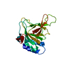
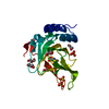
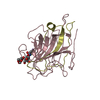
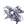
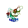
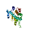
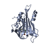
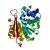
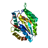
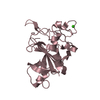
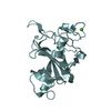
SHEET THE SHEET STRUCTURE OF THIS MOLECULE IS BIFURCATED. IN ORDER TO REPRESENT THIS FEATURE IN ... SHEET THE SHEET STRUCTURE OF THIS MOLECULE IS BIFURCATED. IN ORDER TO REPRESENT THIS FEATURE IN THE SHEET RECORDS BELOW, TWO SHEETS ARE DEFINED.

-
Structure visualization

Structure viewerMolecule:
MolmilJmol/JSmol

Downloads & links

-
Assembly

Deposited unit
C: FICOLIN-1
F: FICOLIN-1
hetero molecules


Theoretical massNumber of molelcules
Total (without water)48,8324
Polymers48,7522
Non-polymers802
Water4,594255
1
C: FICOLIN-1
hetero molecules


Theoretical massNumber of molelcules
Total (without water)24,4162
Polymers24,3761
Non-polymers401
Water181
TypeNameSymmetry operationNumber
identity operation1_555x,y,z1
MethodPQS
2
F: FICOLIN-1
hetero molecules


Theoretical massNumber of molelcules
Total (without water)24,4162
Polymers24,3761
Non-polymers401
Water181
TypeNameSymmetry operationNumber
identity operation1_555x,y,z1
MethodPQS
Unit cell
Length a, b, c (Å)69.400, 69.400, 77.630
Angle α, β, γ (deg.)90.00, 90.00, 120.00
Int Tables number143
Space group name H-MP3

-
Components

#1: Protein FICOLIN-1 / FICOLIN-A / FICOLIN-ALPHA / M-FICOLIN / COLLAGEN/ FIBRINOGEN DOMAIN-CONTAINING PROTEIN 1


Mass: 24375.922 Da / Num. of mol.: 2 / Fragment: C-TERMINAL DOMAIN, RESIDUES 109-326
Source method: isolated from a genetically manipulated source
Source: (gene. exp.) HOMO SAPIENS (human) / Cell line (production host): High Five / Production host: TRICHOPLUSIA NI (cabbage looper) / References: UniProt: O00602
#2: Chemical ChemComp-CA / CALCIUM ION


Mass: 40.078 Da / Num. of mol.: 2 / Source method: obtained synthetically / Formula: Ca
#3: Water ChemComp-HOH / water


Mass: 18.015 Da / Num. of mol.: 255 / Source method: isolated from a natural source / Formula: H2O

-
Experimental details

-
Experiment

ExperimentMethod: X-RAY DIFFRACTION / Number of used crystals: 1

-
Sample preparation

CrystalDensity Matthews: 2.07 Å3/Da / Density % sol: 40.06 % / Description: NONE
Crystal growTemperature: 293 K / Method: vapor diffusion, hanging drop / pH: 5.6 / Details: pH 5.6

-
Data collection

DiffractionMean temperature: 100 K
Diffraction sourceSource: SYNCHROTRON / Site: ESRF / Beamline: ID23-2 / Wavelength: 0.872
DetectorType: MARRESEARCH / Detector: CCD / Date: Dec 16, 2006
RadiationProtocol: SINGLE WAVELENGTH / Monochromatic (M) / Laue (L): M / Scattering type: x-ray
Radiation wavelengthWavelength: 0.872 Å / Relative weight: 1
ReflectionResolution: 1.7→28.03 Å / Num. obs: 45425 / % possible obs: 98.7 % / Observed criterion σ(I): 0 / Redundancy: 5.7 % / Rsym value: 0.073 / Net I/σ(I): 14.9
Reflection shellResolution: 1.7→1.75 Å / Redundancy: 5.7 % / Mean I/σ(I) obs: 3.9 / Rsym value: 0.369 / % possible all: 95.5

-
Processing

Software
NameVersionClassification
REFMAC5.2.0019refinement
XDSdata reduction
XSCALEdata scaling
PHASERphasing
RefinementMethod to determine structure: MOLECULAR REPLACEMENT / Resolution: 1.7→28.03 Å / Cor.coef. Fo:Fc: 0.942 / Cor.coef. Fo:Fc free: 0.923 / SU B: 2.453 / SU ML: 0.084 / Cross valid method: THROUGHOUT / ESU R: 0.134 / ESU R Free: 0.125 / Stereochemistry target values: MAXIMUM LIKELIHOOD
Details: HYDROGENS HAVE BEEN ADDED IN THE RIDING POSITIONS. DISORDERED REGION ARE NOT MODELED
RfactorNum. reflection% reflectionSelection details
Rfree0.252 2301 5 %RANDOM
Rwork0.219 ---
obs0.221 43709 100 %-
Solvent computationIon probe radii: 0.8 Å / Shrinkage radii: 0.8 Å / VDW probe radii: 1.4 Å / Solvent model: MASK
Displacement parametersBiso mean: 22.81 Å2
Baniso -1Baniso -2Baniso -3
1-0.37 Å20.19 Å20 Å2
2--0.37 Å20 Å2
3----0.56 Å2
Refinement stepCycle: LAST / Resolution: 1.7→28.03 Å
ProteinNucleic acidLigandSolventTotal
Num. atoms3288 0 2 255 3545
Refine LS restraints
Refine-IDTypeDev idealDev ideal targetNumber
X-RAY DIFFRACTIONr_bond_refined_d0.0090.0213603
X-RAY DIFFRACTIONr_bond_other_d
X-RAY DIFFRACTIONr_angle_refined_deg1.2711.9134911
X-RAY DIFFRACTIONr_angle_other_deg
X-RAY DIFFRACTIONr_dihedral_angle_1_deg9.6815461
X-RAY DIFFRACTIONr_dihedral_angle_2_deg31.4224.352193
X-RAY DIFFRACTIONr_dihedral_angle_3_deg14.04615569
X-RAY DIFFRACTIONr_dihedral_angle_4_deg14.1221518
X-RAY DIFFRACTIONr_chiral_restr0.1130.2478
X-RAY DIFFRACTIONr_gen_planes_refined0.0040.022896
X-RAY DIFFRACTIONr_gen_planes_other
X-RAY DIFFRACTIONr_nbd_refined0.1890.21509
X-RAY DIFFRACTIONr_nbd_other
X-RAY DIFFRACTIONr_nbtor_refined0.30.22371
X-RAY DIFFRACTIONr_nbtor_other
X-RAY DIFFRACTIONr_xyhbond_nbd_refined0.1220.2159
X-RAY DIFFRACTIONr_xyhbond_nbd_other
X-RAY DIFFRACTIONr_metal_ion_refined
X-RAY DIFFRACTIONr_metal_ion_other
X-RAY DIFFRACTIONr_symmetry_vdw_refined0.2240.266
X-RAY DIFFRACTIONr_symmetry_vdw_other
X-RAY DIFFRACTIONr_symmetry_hbond_refined0.1110.217
X-RAY DIFFRACTIONr_symmetry_hbond_other
X-RAY DIFFRACTIONr_symmetry_metal_ion_refined
X-RAY DIFFRACTIONr_symmetry_metal_ion_other
X-RAY DIFFRACTIONr_mcbond_it0.9121.52238
X-RAY DIFFRACTIONr_mcbond_other
X-RAY DIFFRACTIONr_mcangle_it1.32223453
X-RAY DIFFRACTIONr_mcangle_other
X-RAY DIFFRACTIONr_scbond_it1.66931612
X-RAY DIFFRACTIONr_scbond_other
X-RAY DIFFRACTIONr_scangle_it2.2924.51446
X-RAY DIFFRACTIONr_scangle_other
X-RAY DIFFRACTIONr_long_range_B_refined
X-RAY DIFFRACTIONr_long_range_B_other
X-RAY DIFFRACTIONr_rigid_bond_restr
X-RAY DIFFRACTIONr_sphericity_free
X-RAY DIFFRACTIONr_sphericity_bonded
LS refinement shellResolution: 1.7→1.74 Å / Total num. of bins used: 20 /
RfactorNum. reflection
Rfree0.3 171
Rwork0.26 3244

+
About Yorodumi

-
News

-
Feb 9, 2022. New format data for meta-information of EMDB entries

New format data for meta-information of EMDB entries

  • Version 3 of the EMDB header file is now the official format.
  • The previous official version 1.9 will be removed from the archive.

Related info.:EMDB header

External links:wwPDB to switch to version 3 of the EMDB data model

-
Aug 12, 2020. Covid-19 info

Covid-19 info

URL: https://pdbjlvh1.pdbj.org/emnavi/covid19.php

New page: Covid-19 featured information page in EM Navigator.

Related info.:Covid-19 info / Mar 5, 2020. Novel coronavirus structure data

+
Mar 5, 2020. Novel coronavirus structure data

Novel coronavirus structure data

Related info.:Yorodumi Speices / Aug 12, 2020. Covid-19 info

External links:COVID-19 featured content - PDBj / Molecule of the Month (242):Coronavirus Proteases

+
Jan 31, 2019. EMDB accession codes are about to change! (news from PDBe EMDB page)

EMDB accession codes are about to change! (news from PDBe EMDB page)

  • The allocation of 4 digits for EMDB accession codes will soon come to an end. Whilst these codes will remain in use, new EMDB accession codes will include an additional digit and will expand incrementally as the available range of codes is exhausted. The current 4-digit format prefixed with “EMD-” (i.e. EMD-XXXX) will advance to a 5-digit format (i.e. EMD-XXXXX), and so on. It is currently estimated that the 4-digit codes will be depleted around Spring 2019, at which point the 5-digit format will come into force.
  • The EM Navigator/Yorodumi systems omit the EMD- prefix.

Related info.:Q: What is EMD? / ID/Accession-code notation in Yorodumi/EM Navigator

External links:EMDB Accession Codes are Changing Soon! / Contact to PDBj

+
Jul 12, 2017. Major update of PDB

Major update of PDB

  • wwPDB released updated PDB data conforming to the new PDBx/mmCIF dictionary.
  • This is a major update changing the version number from 4 to 5, and with Remediation, in which all the entries are updated.
  • In this update, many items about electron microscopy experimental information are reorganized (e.g. em_software).
  • Now, EM Navigator and Yorodumi are based on the updated data.

External links:wwPDB Remediation / Enriched Model Files Conforming to OneDep Data Standards Now Available in the PDB FTP Archive

-
Yorodumi

Thousand views of thousand structures

  • Yorodumi is a browser for structure data from EMDB, PDB, SASBDB, etc.
  • This page is also the successor to EM Navigator detail page, and also detail information page/front-end page for Omokage search.
  • The word "yorodu" (or yorozu) is an old Japanese word meaning "ten thousand". "mi" (miru) is to see.

Related info.:EMDB / PDB / SASBDB / Comparison of 3 databanks / Yorodumi Search / Aug 31, 2016. New EM Navigator & Yorodumi / Yorodumi Papers / Jmol/JSmol / Function and homology information / Changes in new EM Navigator and Yorodumi

Read more