 Movie
Movie Controller


+ Open data

- Basic information
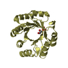
| Entry | Database: PDB / ID: 7p8y | ||||||
|---|---|---|---|---|---|---|---|
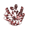
| Title | Short wilavidin apo form | ||||||
 Components Components | Wilavidin | ||||||
 Keywords Keywords | UNKNOWN FUNCTION / Biotin binding protein / avidin / biotechnology | ||||||
| Function / homology | Avidin/streptavidin / Avidin-like superfamily / Avidin family / Avidin-like domain profile. / biotin binding / extracellular region / DI(HYDROXYETHYL)ETHER / TRIETHYLENE GLYCOL / Avidin Function and homology information Function and homology information | ||||||
| Biological species |  Gammaproteobacteria bacterium (bacteria) Gammaproteobacteria bacterium (bacteria) | ||||||
| Method |  X-RAY DIFFRACTION / SYNCHROTRON / MOLECULAR REPLACEMENT / Resolution: 2.18 Å X-RAY DIFFRACTION / SYNCHROTRON / MOLECULAR REPLACEMENT / Resolution: 2.18 Å | ||||||
 Authors Authors | Avraham, O. / Livnah, O. | ||||||
 Citation Citation | Journal: Febs J. / Year: 2022 Title: Wilavidin - a novel member of the avidin family that forms unique biotin-binding hexamers. Authors: Avraham, O. / Bayer, E.A. / Livnah, O. | ||||||
| History |
|
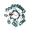
- Structure visualization
| Structure viewer | Molecule: MolmilJmol/JSmol |
|---|
- Downloads & links
-Download
| PDBx/mmCIF format | 7p8y.cif.gz | 101.5 KB | Display | PDBx/mmCIF format |
|---|---|---|---|---|
| PDB format | pdb7p8y.ent.gz | 78.1 KB | Display | PDB format |
| PDBx/mmJSON format | 7p8y.json.gz | Tree view | PDBx/mmJSON format | |
| Others |  Other downloads Other downloads |
-Validation report
| Summary document | 7p8y_validation.pdf.gz | 488.1 KB | Display | wwPDB validaton report |
|---|---|---|---|---|
| Full document | 7p8y_full_validation.pdf.gz | 496.4 KB | Display | |
| Data in XML | 7p8y_validation.xml.gz | 19.9 KB | Display | |
| Data in CIF | 7p8y_validation.cif.gz | 27.9 KB | Display | |
| Arichive directory | https://data.pdbj.org/pub/pdb/validation_reports/p8/7p8yftp://data.pdbj.org/pub/pdb/validation_reports/p8/7p8y | HTTPS FTP |
-Related structure data
| Related structure data |  7ouqSC  7ourC  7p8zC  7ous S: Starting model for refinement C: citing same article ( |
|---|---|
| Similar structure data |


-Links
 PDBj
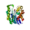
PDBj- Assembly
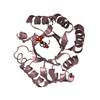
| Deposited unit | 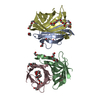
| ||||||||
|---|---|---|---|---|---|---|---|---|---|
| 1 | 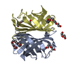
| ||||||||
| 2 | 
| ||||||||
| Unit cell |
|
-Components
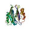
-Protein , 1 types, 4 molecules ABCD
| #1: Protein | Mass: 12786.995 Da / Num. of mol.: 4 Source method: isolated from a genetically manipulated source Details: short wilavidin Source: (gene. exp.) Gammaproteobacteria bacterium (bacteria)Gene: C4528_07355 / Production host: |
|---|
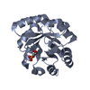
-Non-polymers , 5 types, 181 molecules 


| #2: Chemical | ChemComp-1PE /  Mass: 238.278 Da / Num. of mol.: 6 / Source method: obtained synthetically / Formula: C10H22O6 / Comment: precipitant*YM Mass: 238.278 Da / Num. of mol.: 6 / Source method: obtained synthetically / Formula: C10H22O6 / Comment: precipitant*YM#3: Chemical | ChemComp-P6G /  Mass: 282.331 Da / Num. of mol.: 4 / Source method: obtained synthetically / Formula: C12H26O7 / Comment: precipitant*YM Mass: 282.331 Da / Num. of mol.: 4 / Source method: obtained synthetically / Formula: C12H26O7 / Comment: precipitant*YM#4: Chemical | ChemComp-PEG / |  Mass: 106.120 Da / Num. of mol.: 1 / Source method: obtained synthetically / Formula: C4H10O3 Mass: 106.120 Da / Num. of mol.: 1 / Source method: obtained synthetically / Formula: C4H10O3#5: Chemical | ChemComp-PGE / |  Mass: 150.173 Da / Num. of mol.: 1 / Source method: obtained synthetically / Formula: C6H14O4 Mass: 150.173 Da / Num. of mol.: 1 / Source method: obtained synthetically / Formula: C6H14O4#6: Water | ChemComp-HOH / | Mass: 18.015 Da / Num. of mol.: 169 / Source method: isolated from a natural source / Formula: H2O |
|---|
-Details
| Has ligand of interest | N |
|---|---|
| Has protein modification | Y |
-Experimental details
-Experiment
| Experiment | Method: X-RAY DIFFRACTION / Number of used crystals: 1 |
|---|
- Sample preparation
| Crystal | Density Matthews: 2.3 Å3/Da / Density % sol: 46.51 % |
|---|---|
| Crystal grow | Temperature: 293 K / Method: vapor diffusion, sitting drop Details: 1.5-1.7 M ammonium sulfate 0.1 M HEPES at pH 7.5 2-3% PEG 400 |
-Data collection
| Diffraction | Mean temperature: 100 K / Serial crystal experiment: N |
|---|---|
| Diffraction source | Source: SYNCHROTRON / Site: ESRF  / Beamline: MASSIF-1 / Wavelength: 0.96546 Å / Beamline: MASSIF-1 / Wavelength: 0.96546 Å |
| Detector | Type: DECTRIS PILATUS3 2M / Detector: PIXEL / Date: Dec 2, 2020 |
| Radiation | Protocol: SINGLE WAVELENGTH / Monochromatic (M) / Laue (L): M / Scattering type: x-ray |
| Radiation wavelength | Wavelength: 0.96546 Å / Relative weight: 1 |
| Reflection | Resolution: 2.18→58.71 Å / Num. obs: 25250 / % possible obs: 99.5 % / Redundancy: 4.6 % / CC1/2: 0.993 / Rmerge(I) obs: 0.129 / Net I/σ(I): 9.3 |
| Reflection shell | Resolution: 2.18→2.266 Å / Redundancy: 4.6 % / Num. unique obs: 2805 / CC1/2: 0.984 / % possible all: 99.9 |
- Processing
| Software |
| ||||||||||||||||||||||||||||||||||||||||||||||||||||||||||||
|---|---|---|---|---|---|---|---|---|---|---|---|---|---|---|---|---|---|---|---|---|---|---|---|---|---|---|---|---|---|---|---|---|---|---|---|---|---|---|---|---|---|---|---|---|---|---|---|---|---|---|---|---|---|---|---|---|---|---|---|---|---|
| Refinement | Method to determine structure: MOLECULAR REPLACEMENT Starting model: 7OUQ Resolution: 2.18→58.71 Å / Cor.coef. Fo:Fc: 0.938 / Cor.coef. Fo:Fc free: 0.913 / SU B: 5.962 / SU ML: 0.158 / Cross valid method: THROUGHOUT / σ(F): 0 / ESU R: 0.343 / ESU R Free: 0.264 / Stereochemistry target values: MAXIMUM LIKELIHOOD Details: HYDROGENS HAVE BEEN ADDED IN THE RIDING POSITIONS U VALUES : REFINED INDIVIDUALLY
| ||||||||||||||||||||||||||||||||||||||||||||||||||||||||||||
| Solvent computation | Ion probe radii: 0.8 Å / Shrinkage radii: 0.8 Å / VDW probe radii: 1.2 Å / Solvent model: MASK | ||||||||||||||||||||||||||||||||||||||||||||||||||||||||||||
| Displacement parameters | Biso max: 69.39 Å2 / Biso mean: 18.496 Å2 / Biso min: 7.55 Å2
| ||||||||||||||||||||||||||||||||||||||||||||||||||||||||||||
| Refinement step | Cycle: final / Resolution: 2.18→58.71 Å
| ||||||||||||||||||||||||||||||||||||||||||||||||||||||||||||
| Refine LS restraints |
| ||||||||||||||||||||||||||||||||||||||||||||||||||||||||||||
| LS refinement shell | Resolution: 2.18→2.233 Å / Rfactor Rfree error: 0
|
