- PDB-5cqh: Crystal Structure of the Cancer Genomic DNA Mutator APOBEC3B -
+
データを開く
IDまたはキーワード:
読み込み中...
-
基本情報
登録情報
データベース: PDB / ID: 5cqh
タイトル
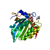
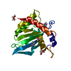
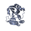
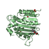
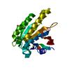
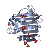
Crystal Structure of the Cancer Genomic DNA Mutator APOBEC3B
要素
DNA dC-dU-editing enzyme APOBEC-3B
キーワード
HYDROLASE / APOBEC / deaminase
機能・相同性
機能・相同性情報
mRNA Editing: C to U Conversion / Formation of the Editosome / single-stranded DNA cytosine deaminase / negative regulation of single stranded viral RNA replication via double stranded DNA intermediate / DNA cytosine deamination / cytidine to uridine editing / clearance of foreign intracellular DNA / cytidine deaminase activity / transposable element silencing / P-body ...mRNA Editing: C to U Conversion / Formation of the Editosome / single-stranded DNA cytosine deaminase / negative regulation of single stranded viral RNA replication via double stranded DNA intermediate / DNA cytosine deamination / cytidine to uridine editing / clearance of foreign intracellular DNA / cytidine deaminase activity / transposable element silencing / P-body / defense response to virus / innate immune response / RNA binding / zinc ion binding / nucleoplasm / nucleus / cytoplasm 類似検索 - 分子機能
: / APOBEC3, cytosine deaminase domain / : / APOBEC/CMP deaminase, zinc-binding / Cytidine and deoxycytidylate deaminases zinc-binding region signature. / Cytidine and deoxycytidylate deaminase domain / Cytidine and deoxycytidylate deaminases domain profile. / Cytidine deaminase-like 類似検索 - ドメイン・相同性
2'-DEOXYCYTIDINE-5'-MONOPHOSPHATE / DNA dC->dU-editing enzyme APOBEC-3B 類似検索 - 構成要素
 ムービー
ムービー コントローラー


 データを開く
 基本情報
基本情報 要素
要素 キーワード
キーワード 機能・相同性情報
 Homo sapiens (ヒト)
 X線回折 /
  データ登録者
 米国, 1件
  引用
引用 構造の表示
構造の表示 ダウンロードとリンク
 その他のダウンロード
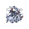


 PDBj
PDBj 集合体
集合体


 分子量: 65.409 Da / 分子数: 1 / 由来タイプ: 合成 / 式: Zn
 タイプ: DNA linking / 分子量: 307.197 Da / 分子数: 1 / 由来タイプ: 合成 / 式: C9H14N3O7P / コメント: dCMP*YM
 分子量: 35.453 Da / 分子数: 1 / 由来タイプ: 合成 / 式: Cl
 分子量: 62.068 Da / 分子数: 4 / 由来タイプ: 合成 / 式: C2H6O2
 試料調製
試料調製 解析
解析