 Movie
Movie Controller

[English] 日本語
 Yorodumi
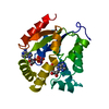
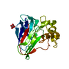
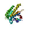
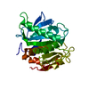
- PDB-6nfm: Crystal Structure of the Cancer Genomic DNA Mutator APOBEC3B with... -
+ Open data

- Basic information
| Entry | Database: PDB / ID: 6nfm | ||||||
|---|---|---|---|---|---|---|---|
| Title | Crystal Structure of the Cancer Genomic DNA Mutator APOBEC3B with loop 7 from APOBEC3G | ||||||
 Components Components | DNA dC->dU-editing enzyme APOBEC-3B | ||||||
 Keywords Keywords | HYDROLASE / APOBEC / deaminase | ||||||
| Function / homology |  Function and homology information Function and homology informationmRNA Editing: C to U Conversion / Formation of the Editosome / single-stranded DNA cytosine deaminase / negative regulation of single stranded viral RNA replication via double stranded DNA intermediate / DNA cytosine deamination / cytidine to uridine editing / clearance of foreign intracellular DNA / cytidine deaminase activity / transposable element silencing / P-body ...mRNA Editing: C to U Conversion / Formation of the Editosome / single-stranded DNA cytosine deaminase / negative regulation of single stranded viral RNA replication via double stranded DNA intermediate / DNA cytosine deamination / cytidine to uridine editing / clearance of foreign intracellular DNA / cytidine deaminase activity / transposable element silencing / P-body / defense response to virus / innate immune response / RNA binding / zinc ion binding / nucleoplasm / nucleus / cytoplasm Similarity search - Function | ||||||
| Biological species |  Homo sapiens (human) Homo sapiens (human) | ||||||
| Method |  X-RAY DIFFRACTION / SYNCHROTRON / MOLECULAR REPLACEMENT / Resolution: 2.53 Å X-RAY DIFFRACTION / SYNCHROTRON / MOLECULAR REPLACEMENT / Resolution: 2.53 Å | ||||||
 Authors Authors | Shi, K. / Aihara, H. | ||||||
| Funding support |  United States, 1items United States, 1items
| ||||||
 Citation Citation | Journal: Faseb Bioadv / Year: 2020 Title: Active site plasticity and possible modes of chemical inhibition of the human DNA deaminase APOBEC3B Authors: Shi, K. / Demir, O. / Carpenter, M.A. / Banerjee, S. / Harki, D.A. / Amaro, R.E. / Harris, R.S. / Aihara, H. | ||||||
| History |
|
- Structure visualization
| Structure viewer | Molecule: MolmilJmol/JSmol |
|---|
- Downloads & links
-Download
| PDBx/mmCIF format | 6nfm.cif.gz | 52.3 KB | Display | PDBx/mmCIF format |
|---|---|---|---|---|
| PDB format | pdb6nfm.ent.gz | 35.3 KB | Display | PDB format |
| PDBx/mmJSON format | 6nfm.json.gz | Tree view | PDBx/mmJSON format | |
| Others |  Other downloads Other downloads |
-Validation report
| Arichive directory | https://data.pdbj.org/pub/pdb/validation_reports/nf/6nfmftp://data.pdbj.org/pub/pdb/validation_reports/nf/6nfm | HTTPS FTP |
|---|
-Related structure data
| Related structure data | 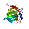 6nfkC 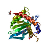 6nflC 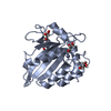 5cqdS S: Starting model for refinement C: citing same article ( |
|---|---|
| Similar structure data |


-Links
 PDBj
PDBj- Assembly
| Deposited unit | 
| ||||||||
|---|---|---|---|---|---|---|---|---|---|
| 1 |
| ||||||||
| Unit cell |
|
-Components
| #1: Protein | Mass: 22844.803 Da / Num. of mol.: 1 Mutation: F200S, W228S, L230K, Y250S, F308K, Y315D, D316Q, P317G, L318R, Y319C, K320Q Source method: isolated from a genetically manipulated source Source: (gene. exp.) Homo sapiens (human) / Gene: APOBEC3B / Production host:  References: UniProt: Q9UH17, single-stranded DNA cytosine deaminase |
|---|---|
| #2: Chemical | ChemComp-CL /   Mass: 35.453 Da / Num. of mol.: 1 / Source method: obtained synthetically / Formula: Cl Mass: 35.453 Da / Num. of mol.: 1 / Source method: obtained synthetically / Formula: Cl |
| #3: Water | ChemComp-HOH /  Mass: 18.015 Da / Num. of mol.: 22 / Source method: isolated from a natural source / Formula: H2O Mass: 18.015 Da / Num. of mol.: 22 / Source method: isolated from a natural source / Formula: H2O |
| Has protein modification | Y |
-Experimental details
-Experiment
| Experiment | Method: X-RAY DIFFRACTION / Number of used crystals: 1 |
|---|
- Sample preparation
| Crystal | Density Matthews: 2.08 Å3/Da / Density % sol: 40.73 % |
|---|---|
| Crystal grow | Temperature: 293 K / Method: vapor diffusion, sitting drop / Details: LiCl, HEPES,PEG3350 |
-Data collection
| Diffraction | Mean temperature: 100 K / Serial crystal experiment: N |
|---|---|
| Diffraction source | Source: SYNCHROTRON / Site: APS / Beamline: 24-ID-E / Wavelength: 0.979 Å |
| Detector | Type: DECTRIS EIGER X 16M / Detector: PIXEL / Date: Feb 26, 2016 |
| Radiation | Protocol: SINGLE WAVELENGTH / Monochromatic (M) / Laue (L): M / Scattering type: x-ray |
| Radiation wavelength | Wavelength: 0.979 Å / Relative weight: 1 |
| Reflection | Resolution: 2.53→41.8 Å / Num. obs: 6968 / % possible obs: 99.9 % / Redundancy: 7.5 % / CC1/2: 0.996 / Rmerge(I) obs: 0.18 / Rpim(I) all: 0.07 / Rrim(I) all: 0.19 / Rsym value: 0.18 / Net I/σ(I): 10.9 |
| Reflection shell | Resolution: 2.53→2.62 Å / Redundancy: 7.8 % / Rmerge(I) obs: 1.9 / Mean I/σ(I) obs: 1.2 / Num. unique obs: 674 / CC1/2: 0.5 / Rpim(I) all: 0.7 / Rrim(I) all: 1.9 / Rsym value: 1.9 / % possible all: 100 |
- Processing
| Software |
| ||||||||||||||||||||||||
|---|---|---|---|---|---|---|---|---|---|---|---|---|---|---|---|---|---|---|---|---|---|---|---|---|---|
| Refinement | Method to determine structure: MOLECULAR REPLACEMENT Starting model: 5CQD Resolution: 2.53→41.766 Å / SU ML: 0.4 / Cross valid method: FREE R-VALUE / σ(F): 1.33 / Phase error: 29.29
| ||||||||||||||||||||||||
| Solvent computation | Shrinkage radii: 0.9 Å / VDW probe radii: 1.11 Å | ||||||||||||||||||||||||
| Refinement step | Cycle: LAST / Resolution: 2.53→41.766 Å
| ||||||||||||||||||||||||
| Refine LS restraints |
| ||||||||||||||||||||||||
| LS refinement shell |
|
