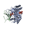
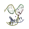
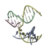
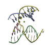
登録情報 データベース : PDB / ID : 7p8kタイトル Crystal structure of in planta processed AvrRps4 in complex with the WRKY domain of RRS1 Avirulence protein,Avirulence protein Disease resistance protein RRS1 キーワード / / / 機能・相同性 分子機能 ドメイン・相同性 構成要素
/ / / / / / / / / / / / / / / / / / / / / / / / / / / / / / / / / / / 生物種 Arabidopsis thaliana (シロイヌナズナ)Pseudomonas syringae (バクテリア)手法 / / / 解像度 : 2.65 Å データ登録者 Mukhi, N. / Brown, H. / Gorenkin, D. / Ding, P. / Bentham, A.R. / Jones, J.D.G. / Banfield, M.J. 資金援助 組織 認可番号 国 European Research Council (ERC) 669926 Biotechnology and Biological Sciences Research Council (BBSRC) BB/M011216/1 Biotechnology and Biological Sciences Research Council (BBSRC) BB/P012574 Biotechnology and Biological Sciences Research Council (BBSRC) BBS/E/J/000PR9795
ジャーナル : Proc.Natl.Acad.Sci.USA / 年 : 2021タイトル : Perception of structurally distinct effectors by the integrated WRKY domain of a plant immune receptor.著者 : Mukhi, N. / Brown, H. / Gorenkin, D. / Ding, P. / Bentham, A.R. / Stevenson, C.E.M. / Jones, J.D.G. / Banfield, M.J. 履歴 登録 2021年7月23日 登録サイト / 処理サイト 改定 1.0 2021年8月4日 Provider / タイプ 改定 1.1 2022年2月2日 Group / カテゴリ / citation_author / database_2Item _citation.country / _citation.journal_abbrev ... _citation.country / _citation.journal_abbrev / _citation.journal_id_ASTM / _citation.journal_id_CSD / _citation.journal_id_ISSN / _citation.journal_volume / _citation.pdbx_database_id_DOI / _citation.pdbx_database_id_PubMed / _citation.title / _citation.year / _database_2.pdbx_DOI / _database_2.pdbx_database_accession 改定 1.2 2024年1月31日 Group / Refinement descriptionカテゴリ / chem_comp_bond / pdbx_initial_refinement_model
すべて表示 表示を減らす
 ムービー
ムービー コントローラー


 データを開く
 基本情報
基本情報 要素
要素 キーワード
キーワード 機能・相同性情報
 Pseudomonas syringae (バクテリア)
 X線回折 /
  データ登録者
 英国, 4件
  引用
引用 構造の表示
構造の表示 ダウンロードとリンク
 その他のダウンロード

 PDBj
PDBj

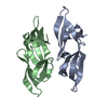
 集合体
集合体

 分子量: 65.409 Da / 分子数: 1 / 由来タイプ: 合成 / 式: Zn / タイプ: SUBJECT OF INVESTIGATION
 分子量: 18.015 Da / 分子数: 3 / 由来タイプ: 天然 / 式: H2O
 試料調製
試料調製 解析
解析