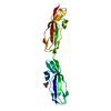
 Movie
Movie Controller

[English] 日本語
 Yorodumi
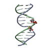
- PDB-7avk: Streptococcal High Identity Repeats in Tandem (SHIRT) domain 10 f... -
+ Open data

- Basic information
| Entry | Database: PDB / ID: 7avk | ||||||
|---|---|---|---|---|---|---|---|
| Title | Streptococcal High Identity Repeats in Tandem (SHIRT) domain 10 from cell surface protein SGO_0707 | ||||||
 Components Components | LPXTG cell wall surface protein | ||||||
 Keywords Keywords | CELL ADHESION / Bacterial surface / adhesin / tandem repeat / Sgo0707 | ||||||
| Function / homology |  Function and homology information Function and homology informationSHIRT domain / Sgo0707, N-terminal domain / SHIRT domain / Sgo0707 N-terminal domain / Sgo0707-like, N2 domain / Sgo0707 N2 domain / Fimbrial isopeptide formation D2 domain / LPXTG cell wall anchor motif / Gram-positive cocci surface proteins LPxTG motif profile. / LPXTG cell wall anchor domain Similarity search - Domain/homology | ||||||
| Biological species |  Streptococcus gordonii (bacteria) Streptococcus gordonii (bacteria) | ||||||
| Method |  X-RAY DIFFRACTION / SYNCHROTRON / MOLECULAR REPLACEMENT / Resolution: 0.82 Å X-RAY DIFFRACTION / SYNCHROTRON / MOLECULAR REPLACEMENT / Resolution: 0.82 Å | ||||||
 Authors Authors | Degut, C. / Gilburt, J. / Whelan, F. / Jenkins, H.T. / Potts, J.R. | ||||||
| Funding support |  United Kingdom, 1items United Kingdom, 1items
| ||||||
 Citation Citation | Journal: Proc.Natl.Acad.Sci.USA / Year: 2021 Title: Periscope Proteins are variable-length regulators of bacterial cell surface interactions. Authors: Whelan, F. / Lafita, A. / Gilburt, J. / Degut, C. / Griffiths, S.C. / Jenkins, H.T. / St John, A.N. / Paci, E. / Moir, J.W.B. / Plevin, M.J. / Baumann, C.G. / Bateman, A. / Potts, J.R. | ||||||
| History |
|
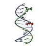
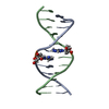
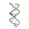
- Structure visualization
| Structure viewer | Molecule: MolmilJmol/JSmol |
|---|
- Downloads & links
-Download
| PDBx/mmCIF format | 7avk.cif.gz | 127.8 KB | Display | PDBx/mmCIF format |
|---|---|---|---|---|
| PDB format | pdb7avk.ent.gz | 102.1 KB | Display | PDB format |
| PDBx/mmJSON format | 7avk.json.gz | Tree view | PDBx/mmJSON format | |
| Others |  Other downloads Other downloads |
-Validation report
| Arichive directory | https://data.pdbj.org/pub/pdb/validation_reports/av/7avkftp://data.pdbj.org/pub/pdb/validation_reports/av/7avk | HTTPS FTP |
|---|
-Related structure data


-Links
 PDBj
PDBj- Assembly
| Deposited unit | 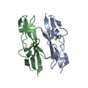
| ||||||||||
|---|---|---|---|---|---|---|---|---|---|---|---|
| 1 | 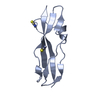
| ||||||||||
| 2 | 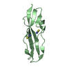
| ||||||||||
| Unit cell |
| ||||||||||
| Components on special symmetry positions |
|
-Components
| #1: Protein | Mass: 9560.514 Da / Num. of mol.: 2 Source method: isolated from a genetically manipulated source Source: (gene. exp.) Streptococcus gordonii (strain Challis / ATCC 35105 / BCRC 15272 / CH1 / DL1 / V288) (bacteria)Strain: Challis / ATCC 35105 / BCRC 15272 / CH1 / DL1 / V288 Gene: SGO_0707 / Production host: #2: Chemical | ChemComp-IS8 /   Mass: 59.090 Da / Num. of mol.: 5 / Source method: obtained synthetically / Formula: CHNS Mass: 59.090 Da / Num. of mol.: 5 / Source method: obtained synthetically / Formula: CHNS#3: Water | ChemComp-HOH / |  Mass: 18.015 Da / Num. of mol.: 235 / Source method: isolated from a natural source / Formula: H2O Mass: 18.015 Da / Num. of mol.: 235 / Source method: isolated from a natural source / Formula: H2OHas ligand of interest | N | |
|---|
-Experimental details
-Experiment
| Experiment | Method: X-RAY DIFFRACTION / Number of used crystals: 1 |
|---|
- Sample preparation
| Crystal | Density Matthews: 2.07 Å3/Da / Density % sol: 40.68 % |
|---|---|
| Crystal grow | Temperature: 277 K / Method: vapor diffusion, hanging drop / pH: 7 Details: 2.2 M ammonium sulphate, 150 mM potassium thiocyanate |
-Data collection
| Diffraction | Mean temperature: 100 K / Serial crystal experiment: N |
|---|---|
| Diffraction source | Source: SYNCHROTRON / Site: Diamond / Beamline: I04 / Wavelength: 0.78001 Å |
| Detector | Type: DECTRIS PILATUS3 6M / Detector: PIXEL / Date: Oct 18, 2018 |
| Radiation | Protocol: SINGLE WAVELENGTH / Monochromatic (M) / Laue (L): M / Scattering type: x-ray |
| Radiation wavelength | Wavelength: 0.78001 Å / Relative weight: 1 |
| Reflection | Resolution: 0.82→48.38 Å / Num. obs: 147184 / % possible obs: 96.75 % / Redundancy: 2 % / Biso Wilson estimate: 10.86 Å2 / CC1/2: 1 / CC star: 1 / Rmerge(I) obs: 0.045 / Net I/σ(I): 28.26 |
| Reflection shell | Resolution: 0.82→0.8493 Å / Redundancy: 1.9 % / Rmerge(I) obs: 2.057 / Mean I/σ(I) obs: 0.34 / Num. unique obs: 24481 / CC1/2: 0.239 / CC star: 0.621 / Rrim(I) all: 2.909 / % possible all: 76.5 |
- Processing
| Software |
| |||||||||||||||||||||||||||||||||||||||||||||||||||||||||||||||||||||||||||||
|---|---|---|---|---|---|---|---|---|---|---|---|---|---|---|---|---|---|---|---|---|---|---|---|---|---|---|---|---|---|---|---|---|---|---|---|---|---|---|---|---|---|---|---|---|---|---|---|---|---|---|---|---|---|---|---|---|---|---|---|---|---|---|---|---|---|---|---|---|---|---|---|---|---|---|---|---|---|---|
| Refinement | Method to determine structure: MOLECULAR REPLACEMENT Starting model: SHIRT R2 Resolution: 0.82→48.38 Å / SU ML: 0.17 / Cross valid method: FREE R-VALUE / σ(F): 1.5 / Phase error: 27.03 / Stereochemistry target values: ML
| |||||||||||||||||||||||||||||||||||||||||||||||||||||||||||||||||||||||||||||
| Solvent computation | Shrinkage radii: 0.9 Å / VDW probe radii: 1.11 Å / Solvent model: FLAT BULK SOLVENT MODEL | |||||||||||||||||||||||||||||||||||||||||||||||||||||||||||||||||||||||||||||
| Refinement step | Cycle: LAST / Resolution: 0.82→48.38 Å
| |||||||||||||||||||||||||||||||||||||||||||||||||||||||||||||||||||||||||||||
| Refine LS restraints |
| |||||||||||||||||||||||||||||||||||||||||||||||||||||||||||||||||||||||||||||
| LS refinement shell |
|
