 Movie
Movie Controller


+ Open data

- Basic information
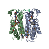
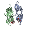
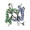
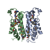
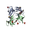
| Entry | Database: PDB / ID: 6x7h | ||||||
|---|---|---|---|---|---|---|---|
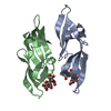
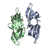
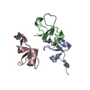
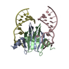
| Title | Cyanovirin-N Mutation I34Y with Dimannose bound | ||||||
 Components Components | Cyanovirin-N | ||||||
 Keywords Keywords | ANTIVIRAL PROTEIN / Mannose binding protein Antiviral protein | ||||||
| Function / homology | Cyanovirin-N / Cyanovirin-N superfamily / CVNH domain / CVNH / regulation of defense response to virus / carbohydrate binding / 2alpha-alpha-mannobiose / Cyanovirin-N Function and homology information Function and homology information | ||||||
| Biological species |  Nostoc ellipsosporum (bacteria) Nostoc ellipsosporum (bacteria) | ||||||
| Method |  X-RAY DIFFRACTION / SYNCHROTRON / MOLECULAR REPLACEMENT / Resolution: 1.25 Å X-RAY DIFFRACTION / SYNCHROTRON / MOLECULAR REPLACEMENT / Resolution: 1.25 Å | ||||||
 Authors Authors | Fromme, R. / Sharma, P. / Ghirlanda, G. | ||||||
 Citation Citation | Journal: Elife / Year: 2022 Title: Design of novel cyanovirin-N variants by modulation of binding dynamics through distal mutations. Authors: Kazan, I.C. / Sharma, P. / Rahman, M.I. / Bobkov, A. / Fromme, R. / Ghirlanda, G. / Ozkan, S.B. #1: Journal: Protein Sci. / Year: 2008Title: Conformational gating of dimannose binding to the antiviral protein cyanovirin revealed from the crystal structure at 1.35 A resolution. Authors: Fromme, R. / Katiliene, Z. / Fromme, P. / Ghirlanda, G. #2: Journal: Biochemistry / Year: 2007Title: A monovalent mutant of cyanovirin-N provides insight into the role of multiple interactions with gp120 for antiviral activity. Authors: Fromme, R. / Katiliene, Z. / Giomarelli, B. / Bogani, F. / Mc Mahon, J. / Mori, T. / Fromme, P. / Ghirlanda, G. | ||||||
| History |
|
- Structure visualization
| Structure viewer | Molecule: MolmilJmol/JSmol |
|---|
- Downloads & links
-Download
| PDBx/mmCIF format | 6x7h.cif.gz | 134.5 KB | Display | PDBx/mmCIF format |
|---|---|---|---|---|
| PDB format | pdb6x7h.ent.gz | 107.2 KB | Display | PDB format |
| PDBx/mmJSON format | 6x7h.json.gz | Tree view | PDBx/mmJSON format | |
| Others |  Other downloads Other downloads |
-Validation report
| Summary document | 6x7h_validation.pdf.gz | 1.2 MB | Display | wwPDB validaton report |
|---|---|---|---|---|
| Full document | 6x7h_full_validation.pdf.gz | 1.2 MB | Display | |
| Data in XML | 6x7h_validation.xml.gz | 13.5 KB | Display | |
| Data in CIF | 6x7h_validation.cif.gz | 19.6 KB | Display | |
| Arichive directory | https://data.pdbj.org/pub/pdb/validation_reports/x7/6x7hftp://data.pdbj.org/pub/pdb/validation_reports/x7/6x7h | HTTPS FTP |
-Related structure data
| Related structure data |  2rdkS S: Starting model for refinement |
|---|---|
| Similar structure data |

-Links
 PDBj
PDBj

- Assembly
| Deposited unit | 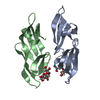
| ||||||||||
|---|---|---|---|---|---|---|---|---|---|---|---|
| 1 |
| ||||||||||
| Unit cell |
|
-Components
| #1: Protein | Mass: 11999.111 Da / Num. of mol.: 2 / Mutation: I34Y Source method: isolated from a genetically manipulated source Source: (gene. exp.) Nostoc ellipsosporum (bacteria) / Production host: #2: Polysaccharide |   Source method: isolated from a genetically manipulated source Details: oligosaccharide / References: 2alpha-alpha-mannobiose #3: Water | ChemComp-HOH / |  Mass: 18.015 Da / Num. of mol.: 296 / Source method: isolated from a natural source / Formula: H2O Mass: 18.015 Da / Num. of mol.: 296 / Source method: isolated from a natural source / Formula: H2OHas ligand of interest | Y | Has protein modification | Y | |
|---|
-Experimental details
-Experiment
| Experiment | Method: X-RAY DIFFRACTION / Number of used crystals: 1 |
|---|
- Sample preparation
| Crystal | Density Matthews: 2.17 Å3/Da / Density % sol: 43.23 % / Description: Needle, 200 by 50 um |
|---|---|
| Crystal grow | Temperature: 298 K / Method: vapor diffusion / Details: 2 M ammonium sulfate, 5% isopropanol |
-Data collection
| Diffraction | Mean temperature: 100 K / Serial crystal experiment: N | ||||||||||||||||||||||||||||||
|---|---|---|---|---|---|---|---|---|---|---|---|---|---|---|---|---|---|---|---|---|---|---|---|---|---|---|---|---|---|---|---|
| Diffraction source | Source: SYNCHROTRON / Site: ALS  / Beamline: 8.2.1 / Wavelength: 0.999 Å / Beamline: 8.2.1 / Wavelength: 0.999 Å | ||||||||||||||||||||||||||||||
| Detector | Type: ADSC QUANTUM 315r / Detector: CCD / Date: Nov 8, 2018 | ||||||||||||||||||||||||||||||
| Radiation | Monochromator: Si (111) / Protocol: SINGLE WAVELENGTH / Monochromatic (M) / Laue (L): M / Scattering type: x-ray | ||||||||||||||||||||||||||||||
| Radiation wavelength | Wavelength: 0.999 Å / Relative weight: 1 | ||||||||||||||||||||||||||||||
| Reflection | Resolution: 1.25→15.43 Å / Num. obs: 52827 / % possible obs: 92.7 % / Redundancy: 3.5 % / CC1/2: 0.991 / Rmerge(I) obs: 0.083 / Rpim(I) all: 0.053 / Rrim(I) all: 0.098 / Net I/σ(I): 9.6 / Num. measured all: 185048 / Scaling rejects: 87 | ||||||||||||||||||||||||||||||
| Reflection shell | Diffraction-ID: 1
|
- Processing
| Software |
| ||||||||||||||||||||||||||||||||||||||||||||||||||||||||||||||||||||||||||||||||||||
|---|---|---|---|---|---|---|---|---|---|---|---|---|---|---|---|---|---|---|---|---|---|---|---|---|---|---|---|---|---|---|---|---|---|---|---|---|---|---|---|---|---|---|---|---|---|---|---|---|---|---|---|---|---|---|---|---|---|---|---|---|---|---|---|---|---|---|---|---|---|---|---|---|---|---|---|---|---|---|---|---|---|---|---|---|---|
| Refinement | Method to determine structure: MOLECULAR REPLACEMENT Starting model: PDB entry 2RDK Resolution: 1.25→15.43 Å / SU ML: 0.1 / Cross valid method: THROUGHOUT / σ(F): 1.35 / Phase error: 15.44 / Stereochemistry target values: ML
| ||||||||||||||||||||||||||||||||||||||||||||||||||||||||||||||||||||||||||||||||||||
| Solvent computation | Shrinkage radii: 0.9 Å / VDW probe radii: 1.11 Å / Solvent model: FLAT BULK SOLVENT MODEL | ||||||||||||||||||||||||||||||||||||||||||||||||||||||||||||||||||||||||||||||||||||
| Displacement parameters | Biso max: 63.07 Å2 / Biso mean: 17.9278 Å2 / Biso min: 8.2 Å2 | ||||||||||||||||||||||||||||||||||||||||||||||||||||||||||||||||||||||||||||||||||||
| Refinement step | Cycle: final / Resolution: 1.25→15.43 Å
| ||||||||||||||||||||||||||||||||||||||||||||||||||||||||||||||||||||||||||||||||||||
| LS refinement shell | Refine-ID: X-RAY DIFFRACTION / Rfactor Rfree error: 0 / Total num. of bins used: 11
|
