 Movie
Movie Controller

[English] 日本語
 Yorodumi
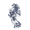
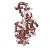
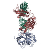
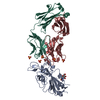
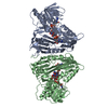
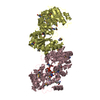
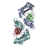
- PDB-7p39: 4,6-alpha-glucanotransferase GtfB from Limosilactobacillus reuter... -
+ Open data

- Basic information
| Entry | Database: PDB / ID: 7p39 | ||||||
|---|---|---|---|---|---|---|---|
| Title | 4,6-alpha-glucanotransferase GtfB from Limosilactobacillus reuteri NCC 2613 complexed with acarbose | ||||||
 Components Components | Dextransucrase | ||||||
 Keywords Keywords | TRANSFERASE / 4 / 6-alpha-transglycosylation / GtfB / Starch conversion | ||||||
| Function / homology |  Function and homology information Function and homology informationdextransucrase activity / dextransucrase / glucan biosynthetic process / glucosyltransferase activity / metal ion binding Similarity search - Function | ||||||
| Biological species |  Lactobacillus reuteri (bacteria) Lactobacillus reuteri (bacteria) | ||||||
| Method |  X-RAY DIFFRACTION / SYNCHROTRON / MOLECULAR REPLACEMENT / molecular replacement / Resolution: 2.9 Å X-RAY DIFFRACTION / SYNCHROTRON / MOLECULAR REPLACEMENT / molecular replacement / Resolution: 2.9 Å | ||||||
 Authors Authors | Pijning, T. / te Poele, E. / Gangoiti, J. / Boerner, T. / Dijkhuizen, L. | ||||||
 Citation Citation | Journal: J.Agric.Food Chem. / Year: 2021 Title: Insights into Broad-Specificity Starch Modification from the Crystal Structure of Limosilactobacillus Reuteri NCC 2613 4,6-alpha-Glucanotransferase GtfB. Authors: Pijning, T. / Gangoiti, J. / Te Poele, E.M. / Borner, T. / Dijkhuizen, L. | ||||||
| History |
|
- Structure visualization
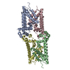
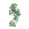
| Structure viewer | Molecule: MolmilJmol/JSmol |
|---|
- Downloads & links
-Download
| PDBx/mmCIF format | 7p39.cif.gz | 335.7 KB | Display | PDBx/mmCIF format |
|---|---|---|---|---|
| PDB format | pdb7p39.ent.gz | 261.9 KB | Display | PDB format |
| PDBx/mmJSON format | 7p39.json.gz | Tree view | PDBx/mmJSON format | |
| Others |  Other downloads Other downloads |
-Validation report
| Arichive directory | https://data.pdbj.org/pub/pdb/validation_reports/p3/7p39ftp://data.pdbj.org/pub/pdb/validation_reports/p3/7p39 | HTTPS FTP |
|---|
-Related structure data

-Links
 PDBj
PDBj
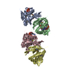
- Assembly
| Deposited unit | 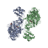
| ||||||||||||||||||
|---|---|---|---|---|---|---|---|---|---|---|---|---|---|---|---|---|---|---|---|
| 1 | 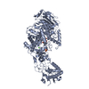
| ||||||||||||||||||
| 2 | 
| ||||||||||||||||||
| Unit cell |
| ||||||||||||||||||
| Noncrystallographic symmetry (NCS) | NCS domain:
NCS domain segments: Component-ID: _ / Ens-ID: 1 / Beg auth comp-ID: VAL / Beg label comp-ID: VAL / End auth comp-ID: ASN / End label comp-ID: ASN / Refine code: _ / Auth seq-ID: 446 - 1277 / Label seq-ID: 50 - 881
|
-Components
| #1: Protein | Mass: 97834.031 Da / Num. of mol.: 2 Source method: isolated from a genetically manipulated source Source: (gene. exp.) Lactobacillus reuteri (bacteria) / Plasmid: pET15b / Production host: #2: Polysaccharide | 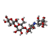  Type: oligosaccharide, Oligosaccharide / Class: Inhibitor / Mass: 645.606 Da / Num. of mol.: 2 Type: oligosaccharide, Oligosaccharide / Class: Inhibitor / Mass: 645.606 Da / Num. of mol.: 2Source method: isolated from a genetically manipulated source Details: oligosaccharide / References: beta-acarbose #3: Chemical |   Mass: 40.078 Da / Num. of mol.: 2 / Source method: obtained synthetically / Formula: Ca Mass: 40.078 Da / Num. of mol.: 2 / Source method: obtained synthetically / Formula: Ca#4: Chemical | ChemComp-SO4 / |   Mass: 96.063 Da / Num. of mol.: 1 / Source method: obtained synthetically / Formula: SO4 Mass: 96.063 Da / Num. of mol.: 1 / Source method: obtained synthetically / Formula: SO4#5: Water | ChemComp-HOH / |  Mass: 18.015 Da / Num. of mol.: 19 / Source method: isolated from a natural source / Formula: H2O Mass: 18.015 Da / Num. of mol.: 19 / Source method: isolated from a natural source / Formula: H2OHas ligand of interest | Y | |
|---|
-Experimental details
-Experiment
| Experiment | Method: X-RAY DIFFRACTION / Number of used crystals: 1 |
|---|
- Sample preparation
| Crystal | Density Matthews: 2.71 Å3/Da / Density % sol: 54.62 % |
|---|---|
| Crystal grow | Temperature: 293 K / Method: vapor diffusion, hanging drop / Details: 1.4-1.7 M (NH4)2SO4, 0.1 M Bis-Tris-HCl, pH 5.5 |
-Data collection
| Diffraction | Mean temperature: 100 K / Serial crystal experiment: N | ||||||||||||||||||||||||||||||
|---|---|---|---|---|---|---|---|---|---|---|---|---|---|---|---|---|---|---|---|---|---|---|---|---|---|---|---|---|---|---|---|
| Diffraction source | Source: SYNCHROTRON / Site: PETRA III, DESY  / Beamline: P11 / Wavelength: 0.9762 Å / Beamline: P11 / Wavelength: 0.9762 Å | ||||||||||||||||||||||||||||||
| Detector | Type: DECTRIS PILATUS 6M-F / Detector: PIXEL / Date: Nov 26, 2017 | ||||||||||||||||||||||||||||||
| Radiation | Protocol: SINGLE WAVELENGTH / Monochromatic (M) / Laue (L): M / Scattering type: x-ray | ||||||||||||||||||||||||||||||
| Radiation wavelength | Wavelength: 0.9762 Å / Relative weight: 1 | ||||||||||||||||||||||||||||||
| Reflection | Resolution: 2.9→49.29 Å / Num. obs: 46821 / % possible obs: 98.3 % / Redundancy: 9.1 % / CC1/2: 0.975 / Rmerge(I) obs: 0.43 / Rpim(I) all: 0.141 / Rrim(I) all: 0.454 / Net I/σ(I): 3.6 | ||||||||||||||||||||||||||||||
| Reflection shell | Diffraction-ID: 1
|
-Phasing
| Phasing | Method: molecular replacement | |||||||||
|---|---|---|---|---|---|---|---|---|---|---|
| Phasing MR | Model details: Phaser MODE: MR_AUTO
|
- Processing
| Software |
| ||||||||||||||||||||||||||||||||||||||||||||||||||||||||||||
|---|---|---|---|---|---|---|---|---|---|---|---|---|---|---|---|---|---|---|---|---|---|---|---|---|---|---|---|---|---|---|---|---|---|---|---|---|---|---|---|---|---|---|---|---|---|---|---|---|---|---|---|---|---|---|---|---|---|---|---|---|---|
| Refinement | Method to determine structure: MOLECULAR REPLACEMENT Starting model: Obtained from Phyre homology modeling Resolution: 2.9→49.29 Å / Cor.coef. Fo:Fc: 0.884 / Cor.coef. Fo:Fc free: 0.857 / SU B: 31.723 / SU ML: 0.535 / Cross valid method: THROUGHOUT / σ(F): 0 / ESU R Free: 0.508 / Stereochemistry target values: MAXIMUM LIKELIHOOD Details: HYDROGENS HAVE BEEN ADDED IN THE RIDING POSITIONS U VALUES : REFINED INDIVIDUALLY
| ||||||||||||||||||||||||||||||||||||||||||||||||||||||||||||
| Solvent computation | Ion probe radii: 0.7 Å / Shrinkage radii: 0.7 Å / VDW probe radii: 1.1 Å / Solvent model: MASK | ||||||||||||||||||||||||||||||||||||||||||||||||||||||||||||
| Displacement parameters | Biso max: 101.97 Å2 / Biso mean: 41.72 Å2 / Biso min: 4.93 Å2
| ||||||||||||||||||||||||||||||||||||||||||||||||||||||||||||
| Refinement step | Cycle: final / Resolution: 2.9→49.29 Å
| ||||||||||||||||||||||||||||||||||||||||||||||||||||||||||||
| Refine LS restraints |
| ||||||||||||||||||||||||||||||||||||||||||||||||||||||||||||
| Refine LS restraints NCS | Ens-ID: 1 / Number: 55432 / Refine-ID: X-RAY DIFFRACTION / Type: interatomic distance / Rms dev position: 0.08 Å / Weight position: 0.05
| ||||||||||||||||||||||||||||||||||||||||||||||||||||||||||||
| LS refinement shell | Resolution: 2.9→2.975 Å / Rfactor Rfree error: 0 / Total num. of bins used: 20
|
