 Movie
Movie Controller


+ Open data

- Basic information
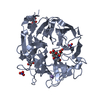
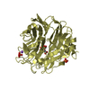
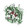
| Entry | Database: PDB / ID: 7p1v | ||||||
|---|---|---|---|---|---|---|---|
| Title | Apo structure of KDNase from Trichophyton Rubrum | ||||||
 Components Components | Extracellular sialidase/neuraminidase | ||||||
 Keywords Keywords | CARBOHYDRATE / Carbohydrate Metabolism / Enzyme Structure / Protein Structure / KDN / KDNase / Sialic Acid / Sialidase | ||||||
| Function / homology |  Function and homology information Function and homology informationganglioside catabolic process / oligosaccharide catabolic process / exo-alpha-sialidase / exo-alpha-sialidase activity / membrane / metal ion binding / cytoplasm Similarity search - Function | ||||||
| Biological species |  Trichophyton rubrum (fungus) Trichophyton rubrum (fungus) | ||||||
| Method |  X-RAY DIFFRACTION / SYNCHROTRON / MOLECULAR REPLACEMENT / Resolution: 1.47 Å X-RAY DIFFRACTION / SYNCHROTRON / MOLECULAR REPLACEMENT / Resolution: 1.47 Å | ||||||
 Authors Authors | Gloster, T.M. / McMahon, S.A. | ||||||
| Funding support | 1items
| ||||||
 Citation Citation | Journal: Acs Chem.Biol. / Year: 2021 Title: Kinetic and Structural Characterization of Sialidases (Kdnases) from Ascomycete Fungal Pathogens. Authors: Nejatie, A. / Steves, E. / Gauthier, N. / Baker, J. / Nesbitt, J. / McMahon, S.A. / Oehler, V. / Thornton, N.J. / Noyovitz, B. / Khazaei, K. / Byers, B.W. / Zandberg, W.F. / Gloster, T.M. / ...Authors: Nejatie, A. / Steves, E. / Gauthier, N. / Baker, J. / Nesbitt, J. / McMahon, S.A. / Oehler, V. / Thornton, N.J. / Noyovitz, B. / Khazaei, K. / Byers, B.W. / Zandberg, W.F. / Gloster, T.M. / Moore, M.M. / Bennet, A.J. | ||||||
| History |
|
- Structure visualization
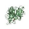
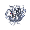
| Structure viewer | Molecule: MolmilJmol/JSmol |
|---|
- Downloads & links
-Download
| PDBx/mmCIF format | 7p1v.cif.gz | 669.1 KB | Display | PDBx/mmCIF format |
|---|---|---|---|---|
| PDB format | pdb7p1v.ent.gz | 551.9 KB | Display | PDB format |
| PDBx/mmJSON format | 7p1v.json.gz | Tree view | PDBx/mmJSON format | |
| Others |  Other downloads Other downloads |
-Validation report
| Arichive directory | https://data.pdbj.org/pub/pdb/validation_reports/p1/7p1vftp://data.pdbj.org/pub/pdb/validation_reports/p1/7p1v | HTTPS FTP |
|---|
-Related structure data
| Related structure data | 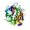 7p1bC 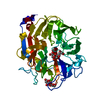 7p1dC 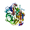 7p1eC 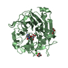 7p1fC 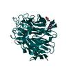 7p1oC  7p1qC 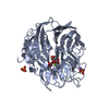 7p1rC 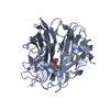 7p1sC 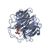 7p1uC 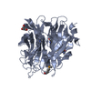 2xcyS S: Starting model for refinement C: citing same article ( |
|---|---|
| Similar structure data |


-Links
 PDBj
PDBj

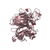
- Assembly
| Deposited unit | 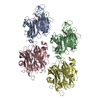
| ||||||||||||||||||||||||||||||||||||||||||||||||||||||||||||||||||||||||||||||||||||||||||||||||||||||||||||||||||||||||||||
|---|---|---|---|---|---|---|---|---|---|---|---|---|---|---|---|---|---|---|---|---|---|---|---|---|---|---|---|---|---|---|---|---|---|---|---|---|---|---|---|---|---|---|---|---|---|---|---|---|---|---|---|---|---|---|---|---|---|---|---|---|---|---|---|---|---|---|---|---|---|---|---|---|---|---|---|---|---|---|---|---|---|---|---|---|---|---|---|---|---|---|---|---|---|---|---|---|---|---|---|---|---|---|---|---|---|---|---|---|---|---|---|---|---|---|---|---|---|---|---|---|---|---|---|---|---|
| 1 | 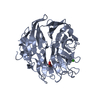
| ||||||||||||||||||||||||||||||||||||||||||||||||||||||||||||||||||||||||||||||||||||||||||||||||||||||||||||||||||||||||||||
| 2 | 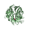
| ||||||||||||||||||||||||||||||||||||||||||||||||||||||||||||||||||||||||||||||||||||||||||||||||||||||||||||||||||||||||||||
| 3 | 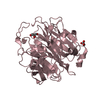
| ||||||||||||||||||||||||||||||||||||||||||||||||||||||||||||||||||||||||||||||||||||||||||||||||||||||||||||||||||||||||||||
| 4 | 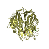
| ||||||||||||||||||||||||||||||||||||||||||||||||||||||||||||||||||||||||||||||||||||||||||||||||||||||||||||||||||||||||||||
| Unit cell |
| ||||||||||||||||||||||||||||||||||||||||||||||||||||||||||||||||||||||||||||||||||||||||||||||||||||||||||||||||||||||||||||
| Noncrystallographic symmetry (NCS) | NCS domain:
NCS domain segments:
NCS ensembles :
|
-Components
| #1: Protein | Mass: 44538.672 Da / Num. of mol.: 4 Source method: isolated from a genetically manipulated source Source: (gene. exp.) Trichophyton rubrum (fungus) / Gene: A7C99_5399 / Production host:  #2: Chemical | ChemComp-GOL /   Mass: 92.094 Da / Num. of mol.: 5 / Source method: obtained synthetically / Formula: C3H8O3 Mass: 92.094 Da / Num. of mol.: 5 / Source method: obtained synthetically / Formula: C3H8O3#3: Chemical | ChemComp-CA /   Mass: 40.078 Da / Num. of mol.: 4 / Source method: isolated from a natural source / Formula: Ca Mass: 40.078 Da / Num. of mol.: 4 / Source method: isolated from a natural source / Formula: Ca#4: Water | ChemComp-HOH / |  Mass: 18.015 Da / Num. of mol.: 1691 / Source method: isolated from a natural source / Formula: H2O Mass: 18.015 Da / Num. of mol.: 1691 / Source method: isolated from a natural source / Formula: H2OHas ligand of interest | N | |
|---|
-Experimental details
-Experiment
| Experiment | Method: X-RAY DIFFRACTION / Number of used crystals: 1 |
|---|
- Sample preparation
| Crystal | Density Matthews: 2.51 Å3/Da / Density % sol: 51 % |
|---|---|
| Crystal grow | Temperature: 293 K / Method: vapor diffusion, sitting drop Details: 0.2 M Calcium chloride dihydrate 0.1 M MES 6.0 20 % w/v PEG 6000 |
-Data collection
| Diffraction | Mean temperature: 173 K / Serial crystal experiment: N |
|---|---|
| Diffraction source | Source: SYNCHROTRON / Site: Diamond  / Beamline: I24 / Wavelength: 0.9688 Å / Beamline: I24 / Wavelength: 0.9688 Å |
| Detector | Type: DECTRIS PILATUS 6M / Detector: PIXEL / Date: Jul 4, 2019 |
| Radiation | Protocol: SINGLE WAVELENGTH / Monochromatic (M) / Laue (L): M / Scattering type: x-ray |
| Radiation wavelength | Wavelength: 0.9688 Å / Relative weight: 1 |
| Reflection | Resolution: 1.47→47.4 Å / Num. obs: 276720 / % possible obs: 98.5 % / Redundancy: 3.2 % / CC1/2: 0.99 / Rmerge(I) obs: 0.066 / Net I/σ(I): 7.3 |
| Reflection shell | Resolution: 1.47→1.51 Å / Rmerge(I) obs: 0.775 / Num. unique obs: 20438 / CC1/2: 0.4 |
- Processing
| Software |
| |||||||||||||||||||||||||||||||||||||||||||||||||||||||||||||||||||||||||||
|---|---|---|---|---|---|---|---|---|---|---|---|---|---|---|---|---|---|---|---|---|---|---|---|---|---|---|---|---|---|---|---|---|---|---|---|---|---|---|---|---|---|---|---|---|---|---|---|---|---|---|---|---|---|---|---|---|---|---|---|---|---|---|---|---|---|---|---|---|---|---|---|---|---|---|---|---|
| Refinement | Method to determine structure: MOLECULAR REPLACEMENT Starting model: 2xcy Resolution: 1.47→47.36 Å / Cor.coef. Fo:Fc: 0.971 / Cor.coef. Fo:Fc free: 0.956 / SU B: 4.665 / SU ML: 0.073 / Cross valid method: THROUGHOUT / σ(F): 0 / ESU R: 0.081 / ESU R Free: 0.076 / Stereochemistry target values: MAXIMUM LIKELIHOOD Details: HYDROGENS HAVE BEEN ADDED IN THE RIDING POSITIONS U VALUES : REFINED INDIVIDUALLY
| |||||||||||||||||||||||||||||||||||||||||||||||||||||||||||||||||||||||||||
| Solvent computation | Ion probe radii: 0.8 Å / Shrinkage radii: 0.8 Å / VDW probe radii: 1.2 Å / Solvent model: MASK | |||||||||||||||||||||||||||||||||||||||||||||||||||||||||||||||||||||||||||
| Displacement parameters | Biso max: 95.18 Å2 / Biso mean: 25.74 Å2 / Biso min: 10.28 Å2
| |||||||||||||||||||||||||||||||||||||||||||||||||||||||||||||||||||||||||||
| Refinement step | Cycle: final / Resolution: 1.47→47.36 Å
| |||||||||||||||||||||||||||||||||||||||||||||||||||||||||||||||||||||||||||
| Refine LS restraints |
| |||||||||||||||||||||||||||||||||||||||||||||||||||||||||||||||||||||||||||
| Refine LS restraints NCS | Refine-ID: X-RAY DIFFRACTION / Type: interatomic distance / Weight position: 0.05
| |||||||||||||||||||||||||||||||||||||||||||||||||||||||||||||||||||||||||||
| LS refinement shell | Resolution: 1.47→1.508 Å / Rfactor Rfree error: 0 / Total num. of bins used: 20
|
