 Movie
Movie Controller

[English] 日本語
 Yorodumi
- PDB-7p1f: Structure of KDNase from Aspergillus terrerus in complex with 2,3... -
+ Open data

- Basic information
| Entry | Database: PDB / ID: 7p1f | |||||||||
|---|---|---|---|---|---|---|---|---|---|---|
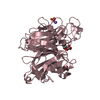
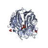
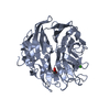
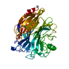
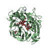
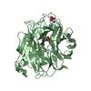
| Title | Structure of KDNase from Aspergillus terrerus in complex with 2,3-didehydro-2,3-dideoxy-D-glycero-D-galacto-nonulosonic acid. | |||||||||
 Components Components | Sialidase domain-containing protein | |||||||||
 Keywords Keywords | CARBOHYDRATE / Carbohydrate Metabolism / Enzyme Structure / Protein Structure / KDN / KDNase / Sialic Acid / Sialidase | |||||||||
| Function / homology |  Function and homology information Function and homology informationganglioside catabolic process / oligosaccharide catabolic process / exo-alpha-sialidase / exo-alpha-sialidase activity / membrane / metal ion binding / cytoplasm Similarity search - Function | |||||||||
| Biological species |  | |||||||||
| Method |  X-RAY DIFFRACTION / SYNCHROTRON / MOLECULAR REPLACEMENT / Resolution: 1.45 Å X-RAY DIFFRACTION / SYNCHROTRON / MOLECULAR REPLACEMENT / Resolution: 1.45 Å | |||||||||
 Authors Authors | Gloster, T.M. / McMahon, S.A. | |||||||||
 Citation Citation | Journal: Acs Chem.Biol. / Year: 2021 Title: Kinetic and Structural Characterization of Sialidases (Kdnases) from Ascomycete Fungal Pathogens. Authors: Nejatie, A. / Steves, E. / Gauthier, N. / Baker, J. / Nesbitt, J. / McMahon, S.A. / Oehler, V. / Thornton, N.J. / Noyovitz, B. / Khazaei, K. / Byers, B.W. / Zandberg, W.F. / Gloster, T.M. / ...Authors: Nejatie, A. / Steves, E. / Gauthier, N. / Baker, J. / Nesbitt, J. / McMahon, S.A. / Oehler, V. / Thornton, N.J. / Noyovitz, B. / Khazaei, K. / Byers, B.W. / Zandberg, W.F. / Gloster, T.M. / Moore, M.M. / Bennet, A.J. | |||||||||
| History |
|
- Structure visualization
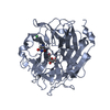
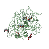
| Structure viewer | Molecule: MolmilJmol/JSmol |
|---|
- Downloads & links
-Download
| PDBx/mmCIF format | 7p1f.cif.gz | 350.5 KB | Display | PDBx/mmCIF format |
|---|---|---|---|---|
| PDB format | pdb7p1f.ent.gz | 280.2 KB | Display | PDB format |
| PDBx/mmJSON format | 7p1f.json.gz | Tree view | PDBx/mmJSON format | |
| Others |  Other downloads Other downloads |
-Validation report
| Arichive directory | https://data.pdbj.org/pub/pdb/validation_reports/p1/7p1fftp://data.pdbj.org/pub/pdb/validation_reports/p1/7p1f | HTTPS FTP |
|---|
-Related structure data
| Related structure data | 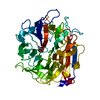 7p1bC 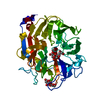 7p1dC 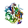 7p1eC 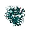 7p1oC 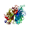 7p1qC  7p1rC 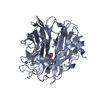 7p1sC 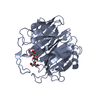 7p1uC  7p1vC 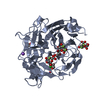 2xzkS S: Starting model for refinement C: citing same article ( |
|---|---|
| Similar structure data |

-Links
 PDBj
PDBj

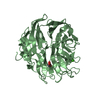
- Assembly
| Deposited unit | 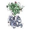
| ||||||||||||||||||||||||||||||||||||||||||||||||
|---|---|---|---|---|---|---|---|---|---|---|---|---|---|---|---|---|---|---|---|---|---|---|---|---|---|---|---|---|---|---|---|---|---|---|---|---|---|---|---|---|---|---|---|---|---|---|---|---|---|
| 1 | 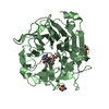
| ||||||||||||||||||||||||||||||||||||||||||||||||
| 2 | 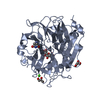
| ||||||||||||||||||||||||||||||||||||||||||||||||
| Unit cell |
| ||||||||||||||||||||||||||||||||||||||||||||||||
| Components on special symmetry positions |
| ||||||||||||||||||||||||||||||||||||||||||||||||
| Noncrystallographic symmetry (NCS) | NCS domain:
NCS domain segments:
NCS ensembles :
|
-Components
| #1: Protein | Mass: 44602.035 Da / Num. of mol.: 2 Source method: isolated from a genetically manipulated source Source: (gene. exp.) Strain: NIH 2624 / FGSC A1156 / Gene: ATEG_04964 / Production host:  #2: Sugar | 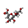  Type: D-saccharide / Mass: 250.203 Da / Num. of mol.: 2 / Source method: isolated from a natural source / Formula: C9H14O8 / Feature type: SUBJECT OF INVESTIGATION Type: D-saccharide / Mass: 250.203 Da / Num. of mol.: 2 / Source method: isolated from a natural source / Formula: C9H14O8 / Feature type: SUBJECT OF INVESTIGATION#3: Chemical | ChemComp-GOL /   Mass: 92.094 Da / Num. of mol.: 4 / Source method: obtained synthetically / Formula: C3H8O3 Mass: 92.094 Da / Num. of mol.: 4 / Source method: obtained synthetically / Formula: C3H8O3#4: Chemical |   Mass: 40.078 Da / Num. of mol.: 2 / Source method: obtained synthetically / Formula: Ca Mass: 40.078 Da / Num. of mol.: 2 / Source method: obtained synthetically / Formula: Ca#5: Water | ChemComp-HOH / |  Mass: 18.015 Da / Num. of mol.: 872 / Source method: isolated from a natural source / Formula: H2O Mass: 18.015 Da / Num. of mol.: 872 / Source method: isolated from a natural source / Formula: H2OHas ligand of interest | Y | Has protein modification | Y | |
|---|
-Experimental details
-Experiment
| Experiment | Method: X-RAY DIFFRACTION / Number of used crystals: 1 |
|---|
- Sample preparation
| Crystal | Density Matthews: 2.27 Å3/Da / Density % sol: 45.82 % |
|---|---|
| Crystal grow | Temperature: 293 K / Method: vapor diffusion, sitting drop / Details: 20% PEG 6K, 0.1M MES pH6 , 0.2M Calcium Chloride |
-Data collection
| Diffraction | Mean temperature: 173 K / Serial crystal experiment: N |
|---|---|
| Diffraction source | Source: SYNCHROTRON / Site: Diamond  / Beamline: I04-1 / Wavelength: 0.9159 Å / Beamline: I04-1 / Wavelength: 0.9159 Å |
| Detector | Type: DECTRIS PILATUS 6M-F / Detector: PIXEL / Date: Dec 16, 2018 |
| Radiation | Protocol: SINGLE WAVELENGTH / Monochromatic (M) / Laue (L): M / Scattering type: x-ray |
| Radiation wavelength | Wavelength: 0.9159 Å / Relative weight: 1 |
| Reflection | Resolution: 1.29→92.8 Å / Num. obs: 129965 / % possible obs: 96.3 % / Redundancy: 2.6 % / CC1/2: 0.996 / Rmerge(I) obs: 0.059 / Net I/σ(I): 7.6 |
| Reflection shell | Resolution: 1.29→1.31 Å / Rmerge(I) obs: 0.68 / Num. unique obs: 8132 / CC1/2: 0.479 / % possible all: 86.6 |
- Processing
| Software |
| |||||||||||||||||||||||||||||||||||||||||||||||||||||||||||||||||||||||||||
|---|---|---|---|---|---|---|---|---|---|---|---|---|---|---|---|---|---|---|---|---|---|---|---|---|---|---|---|---|---|---|---|---|---|---|---|---|---|---|---|---|---|---|---|---|---|---|---|---|---|---|---|---|---|---|---|---|---|---|---|---|---|---|---|---|---|---|---|---|---|---|---|---|---|---|---|---|
| Refinement | Method to determine structure: MOLECULAR REPLACEMENT Starting model: 2xzk Resolution: 1.45→92.8 Å / Cor.coef. Fo:Fc: 0.973 / Cor.coef. Fo:Fc free: 0.956 / SU B: 3.148 / SU ML: 0.052 / Cross valid method: THROUGHOUT / σ(F): 0 / ESU R: 0.076 / ESU R Free: 0.07 / Stereochemistry target values: MAXIMUM LIKELIHOOD Details: HYDROGENS HAVE BEEN ADDED IN THE RIDING POSITIONS U VALUES : REFINED INDIVIDUALLY
| |||||||||||||||||||||||||||||||||||||||||||||||||||||||||||||||||||||||||||
| Solvent computation | Ion probe radii: 0.8 Å / Shrinkage radii: 0.8 Å / VDW probe radii: 1.2 Å / Solvent model: MASK | |||||||||||||||||||||||||||||||||||||||||||||||||||||||||||||||||||||||||||
| Displacement parameters | Biso max: 723.02 Å2 / Biso mean: 17.116 Å2 / Biso min: 5.41 Å2
| |||||||||||||||||||||||||||||||||||||||||||||||||||||||||||||||||||||||||||
| Refinement step | Cycle: final / Resolution: 1.45→92.8 Å
| |||||||||||||||||||||||||||||||||||||||||||||||||||||||||||||||||||||||||||
| Refine LS restraints |
| |||||||||||||||||||||||||||||||||||||||||||||||||||||||||||||||||||||||||||
| Refine LS restraints NCS | Refine-ID: X-RAY DIFFRACTION / Type: interatomic distance / Weight position: 0.05
| |||||||||||||||||||||||||||||||||||||||||||||||||||||||||||||||||||||||||||
| LS refinement shell | Resolution: 1.45→1.488 Å / Rfactor Rfree error: 0 / Total num. of bins used: 20
|
