Mass: 18.015 Da / Num. of mol.: 346 / Source method: isolated from a natural source / Formula: H2O
Has protein modification
Y
Sequence details
THE CONSTRUCT (RESIDUES 24-386) WAS EXPRESSED WITH A PURIFICATION TAG MGSDKIHHHHHHENLYFQG. THE TAG ...THE CONSTRUCT (RESIDUES 24-386) WAS EXPRESSED WITH A PURIFICATION TAG MGSDKIHHHHHHENLYFQG. THE TAG WAS REMOVED WITH TEV PROTEASE LEAVING ONLY A GLYCINE (0) FOLLOWED BY THE TARGET SEQUENCE.
-
Experimental details
-
Experiment
Experiment
Method: X-RAY DIFFRACTION / Number of used crystals: 1
-
Sample preparation
Crystal
Density Matthews: 2.66 Å3/Da / Density % sol: 53.76 %
Crystal grow
Temperature: 277 K / Method: vapor diffusion, sitting drop / pH: 4.2 Details: 40.0% polyethylene glycol 300, 0.1M phosphate-citrate pH 4.2, Additive: 0.003 M fructose, VAPOR DIFFUSION,SITTING DROP,NANODROP, temperature 277K, VAPOR DIFFUSION, SITTING DROP
Resolution: 2.3→28.31 Å / Num. obs: 38497 / % possible obs: 98 % / Observed criterion σ(I): -3 / Redundancy: 5.59 % / Biso Wilson estimate: 37.479 Å2 / Rmerge F obs: 0.259 / Rmerge(I) obs: 0.117 / Rrim(I) all: 0.145 / Net I/σ(I): 8.84 / Num. measured all: 215276
Reflection shell
Diffraction-ID: 1
Resolution (Å)
Highest resolution (Å)
Rmerge F obs
Rmerge(I) obs
Mean I/σ(I) obs
Num. measured obs
Num. possible
Num. unique obs
Rrim(I) all
% possible all
2.3-2.38
1.109
0.705
1.53
18669
7443
6671
0.874
89.6
2.38-2.48
0.935
0.616
1.8
22681
7917
7809
0.761
98.6
2.48-2.59
0.797
0.53
2.2
21391
7445
7349
0.654
98.7
2.59-2.73
0.627
0.401
2.9
22215
7749
7649
0.495
98.7
2.73-2.9
0.421
0.273
4.2
21701
7543
7455
0.337
98.8
2.9-3.12
0.284
0.186
6.1
21563
7486
7417
0.23
99.1
3.12-3.43
0.157
0.108
10.1
21576
7509
7440
0.134
99.1
3.43-3.93
0.091
0.066
15.4
21953
7661
7591
0.082
99.1
3.93-4.93
0.055
0.045
20.8
21481
7522
7462
0.056
99.2
4.93
0.052
0.042
22.5
22046
7743
7650
0.052
98.8
-
Phasing
Phasing
Method: MAD
-
Processing
Software
Name
Version
Classification
NB
SOLVE
phasing
REFMAC
5.5.0109
refinement
XSCALE
datascaling
PDB_EXTRACT
3.1
dataextraction
XDS
datareduction
Refinement
Method to determine structure: MAD / Resolution: 2.3→28.31 Å / Cor.coef. Fo:Fc: 0.948 / Cor.coef. Fo:Fc free: 0.925 / Occupancy max: 1 / Occupancy min: 0.5 / SU B: 11.262 / SU ML: 0.144 / Cross valid method: THROUGHOUT / σ(F): 0 / ESU R: 0.306 / ESU R Free: 0.214 Stereochemistry target values: MAXIMUM LIKELIHOOD WITH PHASES Details: 1. A MET-INHIBITION PROTOCOL WAS USED FOR SELENOMETHIONINE INCORPORATION DURING PROTEIN EXPRESSION. THE OCCUPANCY OF THE SE ATOMS IN THE MSE RESIDUES WAS REDUCED TO 0.75 FOR THE REDUCED ...Details: 1. A MET-INHIBITION PROTOCOL WAS USED FOR SELENOMETHIONINE INCORPORATION DURING PROTEIN EXPRESSION. THE OCCUPANCY OF THE SE ATOMS IN THE MSE RESIDUES WAS REDUCED TO 0.75 FOR THE REDUCED SCATTERING POWER DUE TO PARTIAL S-MET INCORPORATION. 2. POLYEYHYLENE GLYCOL FRAGMENTS FROM THE CRYSTALLIZATION HAVE BEEN MODELED INTO THE STRUCTURE 3.ATOM RECORD CONTAINS SUM OF TLS AND RESIDUAL B FACTORS. ANISOU RECORD CONTAINS SUM OF TLS AND RESIDUAL U FACTORS. 4. WATERS WERE EXCLUDED FROM AUTOMATIC TLS ASSIGNMENT. 5. THE MODELING OF A CIS PEPTIDE BETWEEN GLY 39 AND GLY 40 ON THE A AND B-SUBUNITS IS SUPPORTED BY ELECTRON DENSITY. 6. EVEN THOUGH ASN 77 ON THE B-SUBUNIT IS FLAGGED AS A RAMACHANDRAN OUTLIER IN MOLPROBITY, ITS MODELING IS SUPPORTED BY ELECTRON DENSITY.7. HYDROGENS HAVE BEEN ADDED IN THE RIDING POSITIONS. 8.UNEXPLAINED ELECTRON DENSITIES NEAR THE SIDECHAINS OF LYS 202 ON THE A AND B CHAINS WERE NOT MODELED.
Rfactor
Num. reflection
% reflection
Selection details
Rfree
0.2241
1931
5 %
RANDOM
Rwork
0.1844
36537
-
-
obs
0.1864
38468
99.38 %
-
Solvent computation
Ion probe radii: 0.8 Å / Shrinkage radii: 0.8 Å / VDW probe radii: 1.4 Å / Solvent model: MASK
In the structure databanks used in Yorodumi, some data are registered as the other names, "COVID-19 virus" and "2019-nCoV". Here are the details of the virus and the list of structure data.
Jan 31, 2019. EMDB accession codes are about to change! (news from PDBe EMDB page)
EMDB accession codes are about to change! (news from PDBe EMDB page)
The allocation of 4 digits for EMDB accession codes will soon come to an end. Whilst these codes will remain in use, new EMDB accession codes will include an additional digit and will expand incrementally as the available range of codes is exhausted. The current 4-digit format prefixed with “EMD-” (i.e. EMD-XXXX) will advance to a 5-digit format (i.e. EMD-XXXXX), and so on. It is currently estimated that the 4-digit codes will be depleted around Spring 2019, at which point the 5-digit format will come into force.
The EM Navigator/Yorodumi systems omit the EMD- prefix.
Related info.:Q: What is EMD? / ID/Accession-code notation in Yorodumi/EM Navigator
Yorodumi is a browser for structure data from EMDB, PDB, SASBDB, etc.
This page is also the successor to EM Navigator detail page, and also detail information page/front-end page for Omokage search.
The word "yorodu" (or yorozu) is an old Japanese word meaning "ten thousand". "mi" (miru) is to see.
Related info.:EMDB / PDB / SASBDB / Comparison of 3 databanks / Yorodumi Search / Aug 31, 2016. New EM Navigator & Yorodumi / Yorodumi Papers / Jmol/JSmol / Function and homology information / Changes in new EM Navigator and Yorodumi
 Movie
Movie Controller

 Yorodumi
 Open data
 Basic information
 Components
 Keywords
 Function and homology information
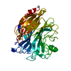
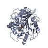
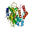
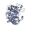
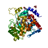
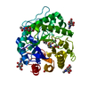
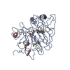
 Bacteroides ovatus (bacteria)
 X-RAY DIFFRACTION /
  Authors
 Citation
 Structure visualization
 Downloads & links
 Other downloads
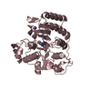


 PDBj
PDBj Assembly

 Mass: 150.173 Da / Num. of mol.: 2 / Source method: obtained synthetically / Formula: C6H14O4
 Mass: 106.120 Da / Num. of mol.: 4 / Source method: obtained synthetically / Formula: C4H10O3
 Mass: 18.015 Da / Num. of mol.: 346 / Source method: isolated from a natural source / Formula: H2O
 Sample preparation
 / Beamline: 8.2.2 / Wavelength: 0.9537,0.9795,0.9793
 Processing