PROTEIN TRANSPORT / Cryo-EM / EspB / ESX-1 / Preferential orientation
Function / homology
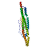
ESX-1 secretion-associated protein EspB, PE domain / : / ESX-1 secreted protein B PE domain / ESX-1 secretion-associated protein EspB, PPE domain / PPE superfamily / extracellular region / ESX-1 secretion-associated protein EspB
Function and homology information
Biological species
Mycobacterium marinum (bacteria)
Method
ELECTRON MICROSCOPY / single particle reconstruction / cryo EM / Resolution: 2.43 Å
Netherlands Organisation for Scientific Research (NWO)
731.016.407
Netherlands
Netherlands Organisation for Scientific Research (NWO)
184.034.014
Netherlands
European Union (EU)
No 766970 Q-SORT
European Union
Consejo Nacional de Ciencia y Tecnologia (CONACYT)
283909
Mexico
Citation
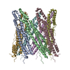
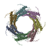
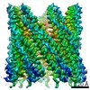
Journal: Curr Res Struct Biol / Year: 2021 Title: Priming mycobacterial ESX-secreted protein B to form a channel-like structure. Authors: Abril Gijsbers / Vanesa Vinciauskaite / Axel Siroy / Ye Gao / Giancarlo Tria / Anjusha Mathew / Nuria Sánchez-Puig / Carmen López-Iglesias / Peter J Peters / Raimond B G Ravelli / Abstract: ESX-1 is a major virulence factor of , a secretion machinery directly involved in the survival of the microorganism from the immune system defence. It disrupts the phagosome membrane of the host cell ...ESX-1 is a major virulence factor of , a secretion machinery directly involved in the survival of the microorganism from the immune system defence. It disrupts the phagosome membrane of the host cell through a contact-dependent mechanism. Recently, the structure of the inner-membrane core complex of the homologous ESX-3 and ESX-5 was resolved; however, the elements involved in the secretion through the outer membrane or those acting on the host cell membrane are unknown. Protein substrates might form this missing element. Here, we describe the oligomerisation process of the ESX-1 substrate EspB, which occurs upon cleavage of its C-terminal region and is favoured by an acidic environment. Cryo-electron microscopy data shows that quaternary structure of EspB is conserved across slow growing species, but not in the fast growing . EspB assembles into a channel with dimensions and characteristics suitable for the transit of ESX-1 substrates, as shown by the presence of another EspB trapped within. Our results provide insight into the structure and assembly of EspB, and suggests a possible function as a structural element of ESX-1.
A: ESX-1 secretion-associated protein EspB B: ESX-1 secretion-associated protein EspB C: ESX-1 secretion-associated protein EspB D: ESX-1 secretion-associated protein EspB E: ESX-1 secretion-associated protein EspB F: ESX-1 secretion-associated protein EspB G: ESX-1 secretion-associated protein EspB
Organism: Escherichia coli (E. coli) / Plasmid: pAG10
Buffer solution
pH: 5.5
Buffer component
ID
Conc.
Name
Formula
Buffer-ID
1
20mM
Acetate
CH3COOH
1
2
150mM
SodiumChloride
NaCl
1
Specimen
Conc.: 8.7 mg/ml / Embedding applied: NO / Shadowing applied: NO / Staining applied: NO / Vitrification applied: YES / Details: This sample was monodisperse.
Instrument: FEI VITROBOT MARK IV / Cryogen name: ETHANE / Humidity: 100 % / Chamber temperature: 277 K
-
Electron microscopy imaging
Experimental equipment
Model: Titan Krios / Image courtesy: FEI Company
Microscopy
Model: TFS KRIOS Details: Basic direct alignments were done as well as astigmatism and coma alignment using AutoCTF
Electron gun
Electron source: FIELD EMISSION GUN / Accelerating voltage: 300 kV / Illumination mode: FLOOD BEAM
Electron lens
Mode: BRIGHT FIELD / Nominal magnification: 105000 X / Calibrated magnification: 105000 X / Nominal defocus max: -2000 nm / Nominal defocus min: -1250 nm / Cs: 2.7 mm / C2 aperture diameter: 50 µm / Alignment procedure: BASIC
Specimen holder
Cryogen: NITROGEN / Specimen holder model: FEI TITAN KRIOS AUTOGRID HOLDER / Temperature (min): 90 K
Image recording
Average exposure time: 1.8 sec. / Electron dose: 40 e/Å2 / Film or detector model: GATAN K3 BIOQUANTUM (6k x 4k) / Num. of grids imaged: 1 / Num. of real images: 2421
EM imaging optics
Energyfilter name: GIF Bioquantum / Energyfilter slit width: 20 eV
Image scans
Sampling size: 5 µm / Width: 5760 / Height: 4092
-
Processing
Software
Name
Version
Classification
phenix.real_space_refine
1.18.2_3874
refinement
PHENIX
1.18.2_3874
refinement
EM software
ID
Name
Version
Category
1
RELION
3.1
particleselection
2
EPU
2.6.1
imageacquisition
4
Gctf
1.06
CTFcorrection
7
Coot
0.9.4
modelfitting
9
RELION
3.1
initialEulerassignment
10
RELION
3.1
finalEulerassignment
11
RELION
3.1
classification
12
RELION
3.1
3Dreconstruction
19
PHENIX
1.18.2
modelrefinement
CTF correction
Type: PHASE FLIPPING AND AMPLITUDE CORRECTION
Symmetry
Point symmetry: C7 (7 fold cyclic)
3D reconstruction
Resolution: 2.43 Å / Resolution method: FSC 0.143 CUT-OFF / Num. of particles: 435505 / Algorithm: FOURIER SPACE / Symmetry type: POINT
Atomic model building
B value: 45.53 / Protocol: OTHER / Space: REAL / Target criteria: Correlation coefficient
In the structure databanks used in Yorodumi, some data are registered as the other names, "COVID-19 virus" and "2019-nCoV". Here are the details of the virus and the list of structure data.
Jan 31, 2019. EMDB accession codes are about to change! (news from PDBe EMDB page)
EMDB accession codes are about to change! (news from PDBe EMDB page)
The allocation of 4 digits for EMDB accession codes will soon come to an end. Whilst these codes will remain in use, new EMDB accession codes will include an additional digit and will expand incrementally as the available range of codes is exhausted. The current 4-digit format prefixed with “EMD-” (i.e. EMD-XXXX) will advance to a 5-digit format (i.e. EMD-XXXXX), and so on. It is currently estimated that the 4-digit codes will be depleted around Spring 2019, at which point the 5-digit format will come into force.
The EM Navigator/Yorodumi systems omit the EMD- prefix.
Related info.:Q: What is EMD? / ID/Accession-code notation in Yorodumi/EM Navigator
Yorodumi is a browser for structure data from EMDB, PDB, SASBDB, etc.
This page is also the successor to EM Navigator detail page, and also detail information page/front-end page for Omokage search.
The word "yorodu" (or yorozu) is an old Japanese word meaning "ten thousand". "mi" (miru) is to see.
Related info.:EMDB / PDB / SASBDB / Comparison of 3 databanks / Yorodumi Search / Aug 31, 2016. New EM Navigator & Yorodumi / Yorodumi Papers / Jmol/JSmol / Function and homology information / Changes in new EM Navigator and Yorodumi
 Movie
Movie Controller


 Open data
 Basic information
 Components
 Keywords
 Function and homology information
 Mycobacterium marinum (bacteria)
 Authors
 Netherlands, European Union,
  Mexico, 4items
  Citation
 Structure visualization
 Downloads & links
 Other downloads
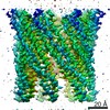


 PDBj
PDBj Assembly
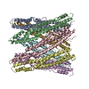
 Sample preparation
 Electron microscopy imaging
 FIELD EMISSION GUN / Accelerating voltage: 300 kV / Illumination mode: FLOOD BEAM
 Processing