 Movie
Movie Controller


+ Open data

- Basic information
| Entry | Database: PDB / ID: 7p02 | |||||||||||||||||||||||||||
|---|---|---|---|---|---|---|---|---|---|---|---|---|---|---|---|---|---|---|---|---|---|---|---|---|---|---|---|---|
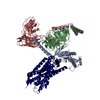
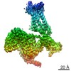
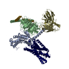
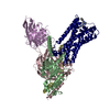
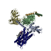
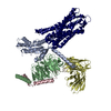
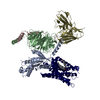
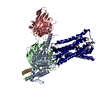
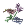
| Title | Human Neurokinin 1 receptor (NK1R) substance P Gs complex | |||||||||||||||||||||||||||
 Components Components |
| |||||||||||||||||||||||||||
 Keywords Keywords | MEMBRANE PROTEIN / Receptor / Complex / Eukaryotic protein | |||||||||||||||||||||||||||
| Function / homology |  Function and homology information Function and homology informationsubstance P receptor activity / substance P receptor binding / positive regulation of corticosterone secretion / aggressive behavior / tachykinin receptor activity / phospholipase C-activating tachykinin receptor signaling pathway / positive regulation of flagellated sperm motility / Tachykinin receptors bind tachykinins / insemination / positive regulation of uterine smooth muscle contraction ...substance P receptor activity / substance P receptor binding / positive regulation of corticosterone secretion / aggressive behavior / tachykinin receptor activity / phospholipase C-activating tachykinin receptor signaling pathway / positive regulation of flagellated sperm motility / Tachykinin receptors bind tachykinins / insemination / positive regulation of uterine smooth muscle contraction / positive regulation of synaptic transmission, cholinergic / detection of abiotic stimulus / sperm ejaculation / positive regulation of lymphocyte proliferation / tachykinin receptor signaling pathway / response to ozone / sperm head / operant conditioning / positive regulation of hormone secretion / positive regulation of action potential / positive regulation of acute inflammatory response / positive regulation of blood pressure / response to auditory stimulus / smooth muscle contraction involved in micturition / regulation of smooth muscle cell proliferation / positive regulation of vascular permeability / positive regulation of leukocyte migration / regulation of smooth muscle cell migration / positive regulation of ossification / response to pain / negative regulation of heart rate / eating behavior / adenylate cyclase-activating G protein-coupled bile acid receptor signaling pathway / adenylate cyclase-activating serotonin receptor signaling pathway / angiotensin-mediated drinking behavior / positive regulation of epithelial cell migration / regulation of skeletal muscle contraction / associative learning / behavioral response to pain / PKA activation in glucagon signalling / response to electrical stimulus / hair follicle placode formation / developmental growth / positive regulation of vasoconstriction / intracellular transport / D1 dopamine receptor binding / sperm flagellum / neuronal dense core vesicle / long-term memory / vascular endothelial cell response to laminar fluid shear stress / renal water homeostasis / Hedgehog 'off' state / activation of adenylate cyclase activity / adenylate cyclase-activating adrenergic receptor signaling pathway / cellular response to acidic pH / positive regulation of stress fiber assembly / regulation of eating behavior / response to progesterone / adenylate cyclase inhibitor activity / response to hormone / positive regulation of protein localization to cell cortex / T cell migration / positive regulation of relaxation of smooth muscle / Adenylate cyclase inhibitory pathway / cellular response to glucagon stimulus / D2 dopamine receptor binding / positive regulation of epithelial cell proliferation / sensory perception of pain / adenylate cyclase-inhibiting serotonin receptor signaling pathway / G protein-coupled serotonin receptor binding / intracellular glucose homeostasis / positive regulation of synaptic transmission, GABAergic / positive regulation of insulin secretion involved in cellular response to glucose stimulus / cellular response to forskolin / adenylate cyclase activator activity / trans-Golgi network membrane / mast cell degranulation / regulation of mitotic spindle organization / chemokine-mediated signaling pathway / response to nicotine / negative regulation of inflammatory response to antigenic stimulus / Regulation of insulin secretion / neuropeptide signaling pathway / response to prostaglandin E / cellular response to nerve growth factor stimulus / bone development / positive regulation of cholesterol biosynthetic process / platelet aggregation / G protein-coupled receptor binding / regulation of blood pressure / response to peptide hormone / cognition / G-protein beta/gamma-subunit complex binding / adenylate cyclase-modulating G protein-coupled receptor signaling pathway / positive regulation of insulin secretion / adenylate cyclase-inhibiting G protein-coupled receptor signaling pathway / Olfactory Signaling Pathway / Activation of the phototransduction cascade / G protein-coupled acetylcholine receptor signaling pathway / G beta:gamma signalling through PLC beta Similarity search - Function | |||||||||||||||||||||||||||
| Biological species |   Homo sapiens (human) Homo sapiens (human) | |||||||||||||||||||||||||||
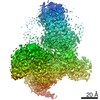
| Method | ELECTRON MICROSCOPY / single particle reconstruction / cryo EM / Resolution: 2.87 Å | |||||||||||||||||||||||||||
 Authors Authors | Thom, C. / Ehrenmann, J. / Vacca, S. / Waltenspuhl, Y. / Schoppe, J. / Medalia, O. / Pluckthun, A. | |||||||||||||||||||||||||||
| Funding support |  Switzerland, 2items Switzerland, 2items
| |||||||||||||||||||||||||||
 Citation Citation | Journal: Sci Adv / Year: 2021 Title: Structures of neurokinin 1 receptor in complex with G and G proteins reveal substance P binding mode and unique activation features. Authors: Cristian Thom / Janosch Ehrenmann / Santiago Vacca / Yann Waltenspühl / Jendrik Schöppe / Ohad Medalia / Andreas Plückthun / Abstract: The neurokinin 1 receptor (NKR) is involved in inflammation and pain transmission. This pathophysiologically important G protein–coupled receptor is predominantly activated by its cognate agonist ...The neurokinin 1 receptor (NKR) is involved in inflammation and pain transmission. This pathophysiologically important G protein–coupled receptor is predominantly activated by its cognate agonist substance P (SP) but also by the closely related neurokinins A and B. Here, we report cryo–electron microscopy structures of SP-bound NKR in complex with its primary downstream signal mediators, G and G. Our structures reveal how a polar network at the extracellular, solvent-exposed receptor surface shapes the orthosteric pocket and that NKR adopts a noncanonical active-state conformation with an interface for G protein binding, which is distinct from previously reported structures. Detailed comparisons with antagonist-bound NKR crystal structures reveal that insurmountable antagonists induce a distinct and long-lasting receptor conformation that sterically blocks SP binding. Together, our structures provide important structural insights into ligand and G protein promiscuity, the lack of basal signaling, and agonist- and antagonist-induced conformations in the neurokinin receptor family. | |||||||||||||||||||||||||||
| History |
|
- Structure visualization
| Movie |
Movie viewer |
|---|---|
| Structure viewer | Molecule: MolmilJmol/JSmol |
- Downloads & links
-Download
| PDBx/mmCIF format | 7p02.cif.gz | 220.6 KB | Display | PDBx/mmCIF format |
|---|---|---|---|---|
| PDB format | pdb7p02.ent.gz | 168.7 KB | Display | PDB format |
| PDBx/mmJSON format | 7p02.json.gz | Tree view | PDBx/mmJSON format | |
| Others |  Other downloads Other downloads |
-Validation report
| Arichive directory | https://data.pdbj.org/pub/pdb/validation_reports/p0/7p02ftp://data.pdbj.org/pub/pdb/validation_reports/p0/7p02 | HTTPS FTP |
|---|
-Related structure data
| Related structure data | 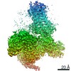 13141MC 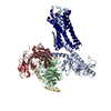 7p00C M: map data used to model this data C: citing same article ( |
|---|---|
| Similar structure data |

-Links
 PDBj
PDBj


- Assembly
| Deposited unit | 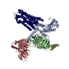
|
|---|---|
| 1 |
|
-Components
-Guanine nucleotide-binding protein ... , 3 types, 3 molecules BGA
| #2: Protein | Mass: 39086.641 Da / Num. of mol.: 1 Source method: isolated from a genetically manipulated source Source: (gene. exp.) Homo sapiens (human) / Gene: GNB1 / Production host:   Spodoptera frugiperda (fall armyworm) / References: UniProt: P62873 Spodoptera frugiperda (fall armyworm) / References: UniProt: P62873 |
|---|---|
| #3: Protein | Mass: 7861.143 Da / Num. of mol.: 1 Source method: isolated from a genetically manipulated source Source: (gene. exp.) Homo sapiens (human) / Gene: GNG2 / Production host: Spodoptera frugiperda (fall armyworm) / References: UniProt: P59768 |
| #4: Protein | Mass: 28428.365 Da / Num. of mol.: 1 Source method: isolated from a genetically manipulated source Source: (gene. exp.) Homo sapiens (human) / Gene: GNAI1, GNAS, GNAS1, GSP / Production host: Spodoptera frugiperda (fall armyworm) / References: UniProt: P63096, UniProt: P63092 |
-Antibody / Protein / Protein/peptide / Non-polymers , 4 types, 4 molecules HRP
| #1: Antibody | Mass: 32409.229 Da / Num. of mol.: 1 Source method: isolated from a genetically manipulated source Source: (gene. exp.) Spodoptera frugiperda (fall armyworm) |
|---|---|
| #5: Protein | Mass: 44218.668 Da / Num. of mol.: 1 Source method: isolated from a genetically manipulated source Source: (gene. exp.) Homo sapiens (human) / Gene: TACR1, NK1R, TAC1R / Production host: Spodoptera frugiperda (fall armyworm) / References: UniProt: P25103 |
| #6: Protein/peptide | Mass: 1348.637 Da / Num. of mol.: 1 Source method: isolated from a genetically manipulated source Source: (gene. exp.) Homo sapiens (human) / Gene: TAC1, NKA, NKNA, TAC2 / Production host: Spodoptera frugiperda (fall armyworm) / References: UniProt: P20366 |
| #7: Chemical | ChemComp-CLR /  Mass: 386.654 Da / Num. of mol.: 1 / Source method: obtained synthetically / Formula: C27H46O Mass: 386.654 Da / Num. of mol.: 1 / Source method: obtained synthetically / Formula: C27H46O |
-Details
| Has ligand of interest | N |
|---|---|
| Has protein modification | Y |
-Experimental details
-Experiment
| Experiment | Method: ELECTRON MICROSCOPY |
|---|---|
| EM experiment | Aggregation state: PARTICLE / 3D reconstruction method: single particle reconstruction |
- Sample preparation
| Component | Name: NK1R in complex with Substance P, heterotrimeric mini-Gs chimera (Gsi) and scFv16 Type: COMPLEX / Entity ID: #1-#6 / Source: RECOMBINANT |
|---|---|
| Source (natural) | Organism: Homo sapiens (human) |
| Source (recombinant) | Organism: Spodoptera frugiperda (fall armyworm) |
| Buffer solution | pH: 7.5 |
| Specimen | Conc.: 1 mg/ml / Embedding applied: NO / Shadowing applied: NO / Staining applied: NO / Vitrification applied: YES |
| Specimen support | Grid material: GOLD / Grid type: Quantifoil R1.2/1.3 |
| Vitrification | Instrument: FEI VITROBOT MARK IV / Cryogen name: ETHANE / Humidity: 100 % / Chamber temperature: 277 K |
- Electron microscopy imaging
| Experimental equipment |  Model: Titan Krios / Image courtesy: FEI Company |
|---|---|
| Microscopy | Model: FEI TITAN KRIOS |
| Electron gun | Electron source:  FIELD EMISSION GUN / Accelerating voltage: 300 kV / Illumination mode: FLOOD BEAM FIELD EMISSION GUN / Accelerating voltage: 300 kV / Illumination mode: FLOOD BEAM |
| Electron lens | Mode: BRIGHT FIELD / Nominal magnification: 130000 X / Nominal defocus max: 2400 nm / Calibrated defocus min: 800 nm / Cs: 2.7 mm |
| Specimen holder | Cryogen: NITROGEN |
| Image recording | Electron dose: 63.51 e/Å2 / Film or detector model: GATAN K3 BIOQUANTUM (6k x 4k) |
- Processing
| Software | Name: PHENIX / Version: 1.19.1_4122: / Classification: refinement | ||||||||||||||||||||||||||||
|---|---|---|---|---|---|---|---|---|---|---|---|---|---|---|---|---|---|---|---|---|---|---|---|---|---|---|---|---|---|
| EM software |
| ||||||||||||||||||||||||||||
| CTF correction | Type: PHASE FLIPPING AND AMPLITUDE CORRECTION | ||||||||||||||||||||||||||||
| Particle selection | Num. of particles selected: 4227825 | ||||||||||||||||||||||||||||
| 3D reconstruction | Resolution: 2.87 Å / Resolution method: FSC 0.143 CUT-OFF / Num. of particles: 395052 / Symmetry type: POINT | ||||||||||||||||||||||||||||
| Refine LS restraints |
|
