 Movie
Movie Controller


+ Open data

- Basic information
| Entry | Database: EMDB / ID: EMD-13141 | |||||||||
|---|---|---|---|---|---|---|---|---|---|---|
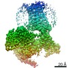
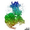
| Title | Human Neurokinin 1 receptor (NK1R) substance P Gs complex | |||||||||
 Map data Map data | processed map | |||||||||
 Sample Sample |
| |||||||||
 Keywords Keywords | Receptor / Complex / Eukaryotic protein / Membrane protein | |||||||||
| Function / homology |  Function and homology information Function and homology informationsubstance P receptor activity / substance P receptor binding / positive regulation of corticosterone secretion / aggressive behavior / tachykinin receptor activity / positive regulation of flagellated sperm motility / insemination / Tachykinin receptors bind tachykinins / phospholipase C-activating tachykinin receptor signaling pathway / positive regulation of uterine smooth muscle contraction ...substance P receptor activity / substance P receptor binding / positive regulation of corticosterone secretion / aggressive behavior / tachykinin receptor activity / positive regulation of flagellated sperm motility / insemination / Tachykinin receptors bind tachykinins / phospholipase C-activating tachykinin receptor signaling pathway / positive regulation of uterine smooth muscle contraction / positive regulation of synaptic transmission, cholinergic / detection of abiotic stimulus / positive regulation of lymphocyte proliferation / sperm ejaculation / tachykinin receptor signaling pathway / response to ozone / sperm head / operant conditioning / positive regulation of hormone secretion / positive regulation of action potential / positive regulation of acute inflammatory response / positive regulation of blood pressure / smooth muscle contraction involved in micturition / response to auditory stimulus / regulation of smooth muscle cell proliferation / positive regulation of leukocyte migration / positive regulation of vascular permeability / regulation of smooth muscle cell migration / positive regulation of ossification / response to pain / negative regulation of heart rate / eating behavior / adenylate cyclase-activating G protein-coupled bile acid receptor signaling pathway / adenylate cyclase-activating serotonin receptor signaling pathway / angiotensin-mediated drinking behavior / positive regulation of epithelial cell migration / associative learning / behavioral response to pain / regulation of skeletal muscle contraction / hair follicle placode formation / PKA activation in glucagon signalling / developmental growth / response to electrical stimulus / positive regulation of vasoconstriction / intracellular transport / D1 dopamine receptor binding / sperm flagellum / long-term memory / neuronal dense core vesicle / renal water homeostasis / vascular endothelial cell response to laminar fluid shear stress / Hedgehog 'off' state / activation of adenylate cyclase activity / positive regulation of stress fiber assembly / adenylate cyclase-activating adrenergic receptor signaling pathway / cellular response to acidic pH / response to progesterone / regulation of eating behavior / adenylate cyclase inhibitor activity / response to hormone / T cell migration / positive regulation of protein localization to cell cortex / positive regulation of relaxation of smooth muscle / Adenylate cyclase inhibitory pathway / sensory perception of pain / cellular response to glucagon stimulus / D2 dopamine receptor binding / positive regulation of epithelial cell proliferation / adenylate cyclase-inhibiting serotonin receptor signaling pathway / G protein-coupled serotonin receptor binding / positive regulation of synaptic transmission, GABAergic / intracellular glucose homeostasis / positive regulation of insulin secretion involved in cellular response to glucose stimulus / adenylate cyclase activator activity / cellular response to forskolin / trans-Golgi network membrane / regulation of mitotic spindle organization / mast cell degranulation / chemokine-mediated signaling pathway / response to nicotine / negative regulation of inflammatory response to antigenic stimulus / neuropeptide signaling pathway / Regulation of insulin secretion / response to prostaglandin E / cellular response to nerve growth factor stimulus / bone development / positive regulation of cholesterol biosynthetic process / platelet aggregation / regulation of blood pressure / G protein-coupled receptor binding / cognition / response to peptide hormone / positive regulation of insulin secretion / G-protein beta/gamma-subunit complex binding / adenylate cyclase-modulating G protein-coupled receptor signaling pathway / adenylate cyclase-inhibiting G protein-coupled receptor signaling pathway / phospholipase C-activating G protein-coupled receptor signaling pathway / Olfactory Signaling Pathway / Activation of the phototransduction cascade / G protein-coupled acetylcholine receptor signaling pathway Similarity search - Function | |||||||||
| Biological species |  Homo sapiens (human) / Homo sapiens (human) /  | |||||||||
| Method | single particle reconstruction / cryo EM / Resolution: 2.87 Å | |||||||||
 Authors Authors | Thom C / Ehrenmann J | |||||||||
| Funding support |  Switzerland, 2 items Switzerland, 2 items
| |||||||||
 Citation Citation | Journal: Sci Adv / Year: 2021 Title: Structures of neurokinin 1 receptor in complex with G and G proteins reveal substance P binding mode and unique activation features. Authors: Cristian Thom / Janosch Ehrenmann / Santiago Vacca / Yann Waltenspühl / Jendrik Schöppe / Ohad Medalia / Andreas Plückthun / Abstract: The neurokinin 1 receptor (NKR) is involved in inflammation and pain transmission. This pathophysiologically important G protein–coupled receptor is predominantly activated by its cognate agonist ...The neurokinin 1 receptor (NKR) is involved in inflammation and pain transmission. This pathophysiologically important G protein–coupled receptor is predominantly activated by its cognate agonist substance P (SP) but also by the closely related neurokinins A and B. Here, we report cryo–electron microscopy structures of SP-bound NKR in complex with its primary downstream signal mediators, G and G. Our structures reveal how a polar network at the extracellular, solvent-exposed receptor surface shapes the orthosteric pocket and that NKR adopts a noncanonical active-state conformation with an interface for G protein binding, which is distinct from previously reported structures. Detailed comparisons with antagonist-bound NKR crystal structures reveal that insurmountable antagonists induce a distinct and long-lasting receptor conformation that sterically blocks SP binding. Together, our structures provide important structural insights into ligand and G protein promiscuity, the lack of basal signaling, and agonist- and antagonist-induced conformations in the neurokinin receptor family. | |||||||||
| History |
|
- Structure visualization
| Movie |
Movie viewer |
|---|---|
| Structure viewer | EM map: SurfViewMolmilJmol/JSmol |
| Supplemental images |

- Downloads & links
-EMDB archive
| Map data | emd_13141.map.gz | 258.7 MB | EMDB map data format | |
|---|---|---|---|---|
| Header (meta data) | emd-13141-v30.xmlemd-13141.xml | 22.6 KB 22.6 KB | Display Display | EMDB header |
| Images | 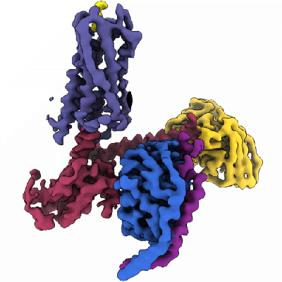 emd_13141.png emd_13141.png | 46.9 KB | ||
| Filedesc metadata | emd-13141.cif.gz | 7 KB | ||
| Others | emd_13141_additional_1.map.gz | 134.9 MB | ||
| Archive directory |  http://ftp.pdbj.org/pub/emdb/structures/EMD-13141ftp://ftp.pdbj.org/pub/emdb/structures/EMD-13141 http://ftp.pdbj.org/pub/emdb/structures/EMD-13141ftp://ftp.pdbj.org/pub/emdb/structures/EMD-13141 | HTTPS FTP |
-Related structure data
| Related structure data |  7p02MC  7p00C M: atomic model generated by this map C: citing same article ( |
|---|---|
| Similar structure data |


-Links
| EMDB pages | EMDB (EBI/PDBe) / EMDataResource |
|---|---|
| Related items in Molecule of the Month |


-Map
| File | Download / File: emd_13141.map.gz / Format: CCP4 / Size: 274.6 MB / Type: IMAGE STORED AS FLOATING POINT NUMBER (4 BYTES) | ||||||||||||||||||||||||||||||||||||||||||||||||||||||||||||
|---|---|---|---|---|---|---|---|---|---|---|---|---|---|---|---|---|---|---|---|---|---|---|---|---|---|---|---|---|---|---|---|---|---|---|---|---|---|---|---|---|---|---|---|---|---|---|---|---|---|---|---|---|---|---|---|---|---|---|---|---|---|
| Annotation | processed map | ||||||||||||||||||||||||||||||||||||||||||||||||||||||||||||
| Projections & slices | Image control
Images are generated by Spider. | ||||||||||||||||||||||||||||||||||||||||||||||||||||||||||||
| Voxel size | X=Y=Z: 0.651 Å | ||||||||||||||||||||||||||||||||||||||||||||||||||||||||||||
| Density |
| ||||||||||||||||||||||||||||||||||||||||||||||||||||||||||||
| Symmetry | Space group: 1 | ||||||||||||||||||||||||||||||||||||||||||||||||||||||||||||
| Details | EMDB XML:
CCP4 map header:
| ||||||||||||||||||||||||||||||||||||||||||||||||||||||||||||
 Z (Sec.)
 Y (Row.)
 X (Col.)


-Supplemental data
-Additional map: raw map
| File | emd_13141_additional_1.map | ||||||||||||
|---|---|---|---|---|---|---|---|---|---|---|---|---|---|
| Annotation | raw map | ||||||||||||
| Projections & Slices |
| ||||||||||||
| Density Histograms |


- Sample components
-Entire : NK1R in complex with Substance P, heterotrimeric mini-Gs chimera ...
| Entire | Name: NK1R in complex with Substance P, heterotrimeric mini-Gs chimera (Gsi) and scFv16 |
|---|---|
| Components |
|
-Supramolecule #1: NK1R in complex with Substance P, heterotrimeric mini-Gs chimera ...
| Supramolecule | Name: NK1R in complex with Substance P, heterotrimeric mini-Gs chimera (Gsi) and scFv16 type: complex / ID: 1 / Parent: 0 / Macromolecule list: #1-#6 |
|---|---|
| Source (natural) | Organism: Homo sapiens (human) |
-Macromolecule #1: Antibody fragment scFv16
| Macromolecule | Name: Antibody fragment scFv16 / type: protein_or_peptide / ID: 1 / Number of copies: 1 / Enantiomer: LEVO |
|---|---|
| Source (natural) | Organism: |
| Molecular weight | Theoretical: 32.409229 KDa |
| Recombinant expression | Organism:   Spodoptera frugiperda (fall armyworm) Spodoptera frugiperda (fall armyworm) |
| Sequence | String: MKFLVNVALV FMVVYISYIY ADYKDDDDKH HHHHHHHHHL EVLFQGPDVQ LVESGGGLVQ PGGSRKLSCS ASGFAFSSFG MHWVRQAPE KGLEWVAYIS SGSGTIYYAD TVKGRFTISR DDPKNTLFLQ MTSLRSEDTA MYYCVRSIYY YGSSPFDFWG Q GTTLTVSS ...String: MKFLVNVALV FMVVYISYIY ADYKDDDDKH HHHHHHHHHL EVLFQGPDVQ LVESGGGLVQ PGGSRKLSCS ASGFAFSSFG MHWVRQAPE KGLEWVAYIS SGSGTIYYAD TVKGRFTISR DDPKNTLFLQ MTSLRSEDTA MYYCVRSIYY YGSSPFDFWG Q GTTLTVSS GGGGSGGGGS GGGGSDIVMT QATSSVPVTP GESVSISCRS SKSLLHSNGN TYLYWFLQRP GQSPQLLIYR MS NLASGVP DRFSGSGSGT AFTLTISRLE AEDVGVYYCM QHLEYPLTFG AGTKLELKAA A |
-Macromolecule #2: Guanine nucleotide-binding protein G(I)/G(S)/G(T) subunit beta-1
| Macromolecule | Name: Guanine nucleotide-binding protein G(I)/G(S)/G(T) subunit beta-1 type: protein_or_peptide / ID: 2 / Number of copies: 1 / Enantiomer: LEVO |
|---|---|
| Source (natural) | Organism: Homo sapiens (human) |
| Molecular weight | Theoretical: 39.086641 KDa |
| Recombinant expression | Organism: Spodoptera frugiperda (fall armyworm) |
| Sequence | String: MHHHHHHHHH HGSSGSELDQ LRQEAEQLKN QIRDARKACA DATLSQITNN IDPVGRIQMR TRRTLRGHLA KIYAMHWGTD SRLLVSASQ DGKLIIWDSY TTNKVHAIPL RSSWVMTCAY APSGNYVACG GLDNICSIYN LKTREGNVRV SRELAGHTGY L SCCRFLDD ...String: MHHHHHHHHH HGSSGSELDQ LRQEAEQLKN QIRDARKACA DATLSQITNN IDPVGRIQMR TRRTLRGHLA KIYAMHWGTD SRLLVSASQ DGKLIIWDSY TTNKVHAIPL RSSWVMTCAY APSGNYVACG GLDNICSIYN LKTREGNVRV SRELAGHTGY L SCCRFLDD NQIVTSSGDT TCALWDIETG QQTTTFTGHT GDVMSLSLAP DTRLFVSGAC DASAKLWDVR EGMCRQTFTG HE SDINAIC FFPNGNAFAT GSDDATCRLF DLRADQELMT YSHDNIICGI TSVSFSKSGR LLLAGYDDFN CNVWDALKAD RAG VLAGHD NRVSCLGVTD DGMAVATGSW DSFLKIWN UniProtKB: Guanine nucleotide-binding protein G(I)/G(S)/G(T) subunit beta-1 |
-Macromolecule #3: Guanine nucleotide-binding protein G(I)/G(S)/G(O) subunit gamma-2
| Macromolecule | Name: Guanine nucleotide-binding protein G(I)/G(S)/G(O) subunit gamma-2 type: protein_or_peptide / ID: 3 / Number of copies: 1 / Enantiomer: LEVO |
|---|---|
| Source (natural) | Organism: Homo sapiens (human) |
| Molecular weight | Theoretical: 7.861143 KDa |
| Recombinant expression | Organism: Spodoptera frugiperda (fall armyworm) |
| Sequence | String: MASNNTASIA QARKLVEQLK MEANIDRIKV SKAAADLMAY CEAHAKEDPL LTPVPASENP FREKKFFCAI L UniProtKB: Guanine nucleotide-binding protein G(I)/G(S)/G(O) subunit gamma-2 |
-Macromolecule #4: Guanine nucleotide-binding protein G(i) subunit alpha-1,Guanine n...
| Macromolecule | Name: Guanine nucleotide-binding protein G(i) subunit alpha-1,Guanine nucleotide-binding protein G(s) subunit alpha isoforms short type: protein_or_peptide / ID: 4 / Number of copies: 1 / Enantiomer: LEVO |
|---|---|
| Source (natural) | Organism: Homo sapiens (human) |
| Molecular weight | Theoretical: 28.428365 KDa |
| Recombinant expression | Organism: Spodoptera frugiperda (fall armyworm) |
| Sequence | String: MGCTLSAEDK AAVERSKMIE KQLQKDKQVY RATHRLLLLG ADNSGKSTIV KQMRILHGGS GGSGGTSGIF ETKFQVDKVN FHMFDVGGQ RDERRKWIQC FNDVTAIIFV VDSSDYNRLQ EALNLFKSIW NNRWLRTISV ILFLNKQDLL AEKVLAGKSK I EDYFPEFA ...String: MGCTLSAEDK AAVERSKMIE KQLQKDKQVY RATHRLLLLG ADNSGKSTIV KQMRILHGGS GGSGGTSGIF ETKFQVDKVN FHMFDVGGQ RDERRKWIQC FNDVTAIIFV VDSSDYNRLQ EALNLFKSIW NNRWLRTISV ILFLNKQDLL AEKVLAGKSK I EDYFPEFA RYTTPEDATP EPGEDPRVTR AKYFIRDEFL RISTASGDGR HYCYPHFTCA VDTENARRIF NDCRDIIQRM HL RQYELL UniProtKB: Guanine nucleotide-binding protein G(i) subunit alpha-1, Guanine nucleotide-binding protein G(s) subunit alpha isoforms short |
-Macromolecule #5: Substance-P receptor
| Macromolecule | Name: Substance-P receptor / type: protein_or_peptide / ID: 5 / Number of copies: 1 / Enantiomer: LEVO |
|---|---|
| Source (natural) | Organism: Homo sapiens (human) |
| Molecular weight | Theoretical: 44.218668 KDa |
| Recombinant expression | Organism: Spodoptera frugiperda (fall armyworm) |
| Sequence | String: MKFLVNVALV FMVVYISYIY ADYKDDDDKH HHHHHHHHHL EVLFQGPMDN VLPVDSDLSP NISTNTSEPN QFVQPAWQIV LWAAAYTVI VVTSVVGNVV VMWIILAHKR MRTVTNYFLV NLAFAEASMA AFNTVVNFTY AVHNEWYYGL FYCKFHNFFP I AAVFASIY ...String: MKFLVNVALV FMVVYISYIY ADYKDDDDKH HHHHHHHHHL EVLFQGPMDN VLPVDSDLSP NISTNTSEPN QFVQPAWQIV LWAAAYTVI VVTSVVGNVV VMWIILAHKR MRTVTNYFLV NLAFAEASMA AFNTVVNFTY AVHNEWYYGL FYCKFHNFFP I AAVFASIY SMTAVAFDRY MAIIHPLQPR LSATATKVVI CVIWVLALLL AFPQGYYSTT ETMPSRVVCM IEWPEHPNKI YE KVYHICV TVLIYFLPLL VIGYAYTVVG ITLWASEIPG DSSDRYHEQV SAKRKVVKMM IVVVCTFAIC WLPFHIFFLL PYI NPDLYL KKFIQQVYLA IMWLAMSSTM YNPIIYCCLN DRFRLGFKHA FRCCPFISAG DYEGLE UniProtKB: Substance-P receptor |
-Macromolecule #6: Protachykinin-1
| Macromolecule | Name: Protachykinin-1 / type: protein_or_peptide / ID: 6 / Number of copies: 1 / Enantiomer: LEVO |
|---|---|
| Source (natural) | Organism: Homo sapiens (human) |
| Molecular weight | Theoretical: 1.348637 KDa |
| Recombinant expression | Organism: Spodoptera frugiperda (fall armyworm) |
| Sequence | String: RPKPQQFFGL M(NH2) UniProtKB: Protachykinin-1 |
-Macromolecule #7: CHOLESTEROL
| Macromolecule | Name: CHOLESTEROL / type: ligand / ID: 7 / Number of copies: 1 / Formula: CLR |
|---|---|
| Molecular weight | Theoretical: 386.654 Da |
| Chemical component information |  ChemComp-CLR: |
-Experimental details
-Structure determination
| Method | cryo EM |
|---|---|
 Processing Processing | single particle reconstruction |
| Aggregation state | particle |
-Sample preparation
| Concentration | 1 mg/mL |
|---|---|
| Buffer | pH: 7.5 |
| Grid | Model: Quantifoil R1.2/1.3 / Material: GOLD / Support film - Material: CARBON / Support film - topology: HOLEY |
| Vitrification | Cryogen name: ETHANE / Chamber humidity: 100 % / Chamber temperature: 277 K / Instrument: FEI VITROBOT MARK IV |
- Electron microscopy
| Microscope | FEI TITAN KRIOS |
|---|---|
| Image recording | Film or detector model: GATAN K3 BIOQUANTUM (6k x 4k) / Average electron dose: 63.51 e/Å2 |
| Electron beam | Acceleration voltage: 300 kV / Electron source:  FIELD EMISSION GUN FIELD EMISSION GUN |
| Electron optics | Calibrated defocus min: 0.8 µm / Illumination mode: FLOOD BEAM / Imaging mode: BRIGHT FIELD / Cs: 2.7 mm / Nominal defocus max: 2.4 µm / Nominal magnification: 130000 |
| Sample stage | Cooling holder cryogen: NITROGEN |
| Experimental equipment |  Model: Titan Krios / Image courtesy: FEI Company |
