 Movie
Movie Controller

[English] 日本語
 Yorodumi
- PDB-7oub: High resolution structure of Alpha-1-acid glycoprotein bound to p... -
+ Open data

- Basic information
| Entry | Database: PDB / ID: 7oub | |||||||||
|---|---|---|---|---|---|---|---|---|---|---|
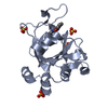
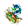
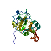
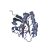
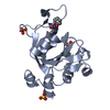
| Title | High resolution structure of Alpha-1-acid glycoprotein bound to potent anti-tumour compound UCN-01 | |||||||||
 Components Components | Alpha-1-acid glycoprotein 2 | |||||||||
 Keywords Keywords | PROTEIN TRANSPORT / AGP2 / ORM2 / lipocalin / complex / Alpha-1-acid Glycoprotein / UCN-01 | |||||||||
| Function / homology |  Function and homology information Function and homology informationpositive regulation of interleukin-1 production / regulation of immune system process / platelet alpha granule lumen / acute-phase response / positive regulation of interleukin-1 beta production / specific granule lumen / positive regulation of tumor necrosis factor production / azurophil granule lumen / Platelet degranulation / blood microparticle ...positive regulation of interleukin-1 production / regulation of immune system process / platelet alpha granule lumen / acute-phase response / positive regulation of interleukin-1 beta production / specific granule lumen / positive regulation of tumor necrosis factor production / azurophil granule lumen / Platelet degranulation / blood microparticle / Neutrophil degranulation / : / extracellular exosome / extracellular region Similarity search - Function | |||||||||
| Biological species |  Homo sapiens (human) Homo sapiens (human) | |||||||||
| Method |  X-RAY DIFFRACTION / SYNCHROTRON / MOLECULAR REPLACEMENT / Resolution: 1.82 Å X-RAY DIFFRACTION / SYNCHROTRON / MOLECULAR REPLACEMENT / Resolution: 1.82 Å | |||||||||
 Authors Authors | Landin, E.J.B. / Williams, C. / Crump, M.P. | |||||||||
| Funding support |  United Kingdom, 2items United Kingdom, 2items
| |||||||||
 Citation Citation | Journal: J.Biol.Chem. / Year: 2021 Title: The structural basis for high affinity binding of alpha 1-acid glycoprotein to the potent antitumor compound UCN-01. Authors: Landin, E.J.B. / Williams, C. / Ryan, S.A. / Bochel, A. / Akter, N. / Redfield, C. / Sessions, R.B. / Dedi, N. / Taylor, R.J. / Crump, M.P. | |||||||||
| History |
|
- Structure visualization
| Structure viewer | Molecule: MolmilJmol/JSmol |
|---|
- Downloads & links
-Download
| PDBx/mmCIF format | 7oub.cif.gz | 96.4 KB | Display | PDBx/mmCIF format |
|---|---|---|---|---|
| PDB format | pdb7oub.ent.gz | 71.4 KB | Display | PDB format |
| PDBx/mmJSON format | 7oub.json.gz | Tree view | PDBx/mmJSON format | |
| Others |  Other downloads Other downloads |
-Validation report
| Arichive directory | https://data.pdbj.org/pub/pdb/validation_reports/ou/7oubftp://data.pdbj.org/pub/pdb/validation_reports/ou/7oub | HTTPS FTP |
|---|
-Related structure data
| Related structure data |  3apuS S: Starting model for refinement |
|---|---|
| Similar structure data | |
| Other databases |
|


-Links
 PDBj
PDBj

- Assembly
| Deposited unit | 
| ||||||||
|---|---|---|---|---|---|---|---|---|---|
| 1 |
| ||||||||
| Unit cell |
|
-Components
| #1: Protein | Mass: 22544.227 Da / Num. of mol.: 1 / Mutation: C149R Source method: isolated from a genetically manipulated source Details: MAHHHHHHSSGLEVLFQGP are vector derived. During purification the protein is cleaved leaving GP from the vector (-1-0) and the mature native protein starts at the Q (1) of QIP after the ...Details: MAHHHHHHSSGLEVLFQGP are vector derived. During purification the protein is cleaved leaving GP from the vector (-1-0) and the mature native protein starts at the Q (1) of QIP after the cleavage tag. The C-terminal His197 is not visible in the crystal structure. Source: (gene. exp.) Homo sapiens (human) / Gene: ORM2, AGP2 / Plasmid: pOPINF / Production host:  |
|---|---|
| #2: Chemical | ChemComp-UCN / 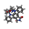  Mass: 482.530 Da / Num. of mol.: 1 / Source method: obtained synthetically / Formula: C28H26N4O4 / Feature type: SUBJECT OF INVESTIGATION Mass: 482.530 Da / Num. of mol.: 1 / Source method: obtained synthetically / Formula: C28H26N4O4 / Feature type: SUBJECT OF INVESTIGATION |
| #3: Water | ChemComp-HOH /  Mass: 18.015 Da / Num. of mol.: 223 / Source method: isolated from a natural source / Formula: H2O Mass: 18.015 Da / Num. of mol.: 223 / Source method: isolated from a natural source / Formula: H2O |
| Has ligand of interest | Y |
| Has protein modification | Y |
-Experimental details
-Experiment
| Experiment | Method: X-RAY DIFFRACTION / Number of used crystals: 1 |
|---|
- Sample preparation
| Crystal | Density Matthews: 2.95 Å3/Da / Density % sol: 58.25 % |
|---|---|
| Crystal grow | Temperature: 294 K / Method: vapor diffusion, sitting drop / pH: 7.4 / Details: 0.1M HEPES 1.4M sodium acetate / PH range: 0.1 |
-Data collection
| Diffraction | Mean temperature: 80 K / Serial crystal experiment: N |
|---|---|
| Diffraction source | Source: SYNCHROTRON / Site: Diamond / Beamline: I24 / Wavelength: 0.9686 Å |
| Detector | Type: DECTRIS PILATUS 6M / Detector: PIXEL / Date: Jan 26, 2020 |
| Radiation | Protocol: SINGLE WAVELENGTH / Monochromatic (M) / Laue (L): M / Scattering type: x-ray |
| Radiation wavelength | Wavelength: 0.9686 Å / Relative weight: 1 |
| Reflection | Resolution: 1.82→76.44 Å / Num. obs: 21541 / % possible obs: 99.68 % / Redundancy: 18.4 % / Biso Wilson estimate: 21.17 Å2 / CC1/2: 0.999 / Rmerge(I) obs: 0.058 / Rpim(I) all: 0.014 / Rrim(I) all: 0.06 / Net I/σ(I): 34.3 |
| Reflection shell | Resolution: 1.82→4.94 Å / Redundancy: 10.3 % / Rmerge(I) obs: 0.16 / Mean I/σ(I) obs: 9.9 / Num. unique obs: 1034 / CC1/2: 0.986 / Rpim(I) all: 0.051 / Rrim(I) all: 0.169 / % possible all: 96 |
- Processing
| Software |
| |||||||||||||||||||||||||||||||||||||||||||||||||||||||||||||||||||||||||||||||||||||||||||||||||||||||||||||||||||||||||||||||||||||||||||||||||||||||||||||||||||||||||||||||||||||||||||||||||||||||||||||||||||||||||||||||||
|---|---|---|---|---|---|---|---|---|---|---|---|---|---|---|---|---|---|---|---|---|---|---|---|---|---|---|---|---|---|---|---|---|---|---|---|---|---|---|---|---|---|---|---|---|---|---|---|---|---|---|---|---|---|---|---|---|---|---|---|---|---|---|---|---|---|---|---|---|---|---|---|---|---|---|---|---|---|---|---|---|---|---|---|---|---|---|---|---|---|---|---|---|---|---|---|---|---|---|---|---|---|---|---|---|---|---|---|---|---|---|---|---|---|---|---|---|---|---|---|---|---|---|---|---|---|---|---|---|---|---|---|---|---|---|---|---|---|---|---|---|---|---|---|---|---|---|---|---|---|---|---|---|---|---|---|---|---|---|---|---|---|---|---|---|---|---|---|---|---|---|---|---|---|---|---|---|---|---|---|---|---|---|---|---|---|---|---|---|---|---|---|---|---|---|---|---|---|---|---|---|---|---|---|---|---|---|---|---|---|---|---|---|---|---|---|---|---|---|---|---|---|---|---|---|---|---|
| Refinement | Method to determine structure: MOLECULAR REPLACEMENT Starting model: 3apu Resolution: 1.82→44.13 Å / SU ML: 0.14 / Cross valid method: THROUGHOUT / σ(F): 1.44 / Phase error: 19.3 / Stereochemistry target values: ML
| |||||||||||||||||||||||||||||||||||||||||||||||||||||||||||||||||||||||||||||||||||||||||||||||||||||||||||||||||||||||||||||||||||||||||||||||||||||||||||||||||||||||||||||||||||||||||||||||||||||||||||||||||||||||||||||||||
| Solvent computation | Shrinkage radii: 0.9 Å / VDW probe radii: 1.11 Å / Solvent model: FLAT BULK SOLVENT MODEL | |||||||||||||||||||||||||||||||||||||||||||||||||||||||||||||||||||||||||||||||||||||||||||||||||||||||||||||||||||||||||||||||||||||||||||||||||||||||||||||||||||||||||||||||||||||||||||||||||||||||||||||||||||||||||||||||||
| Displacement parameters | Biso max: 88.08 Å2 / Biso mean: 30.2026 Å2 / Biso min: 10.62 Å2 | |||||||||||||||||||||||||||||||||||||||||||||||||||||||||||||||||||||||||||||||||||||||||||||||||||||||||||||||||||||||||||||||||||||||||||||||||||||||||||||||||||||||||||||||||||||||||||||||||||||||||||||||||||||||||||||||||
| Refinement step | Cycle: final / Resolution: 1.82→44.13 Å
| |||||||||||||||||||||||||||||||||||||||||||||||||||||||||||||||||||||||||||||||||||||||||||||||||||||||||||||||||||||||||||||||||||||||||||||||||||||||||||||||||||||||||||||||||||||||||||||||||||||||||||||||||||||||||||||||||
| LS refinement shell | Refine-ID: X-RAY DIFFRACTION / Rfactor Rfree error: 0 / Total num. of bins used: 7
| |||||||||||||||||||||||||||||||||||||||||||||||||||||||||||||||||||||||||||||||||||||||||||||||||||||||||||||||||||||||||||||||||||||||||||||||||||||||||||||||||||||||||||||||||||||||||||||||||||||||||||||||||||||||||||||||||
| Refinement TLS params. | Method: refined / Refine-ID: X-RAY DIFFRACTION
| |||||||||||||||||||||||||||||||||||||||||||||||||||||||||||||||||||||||||||||||||||||||||||||||||||||||||||||||||||||||||||||||||||||||||||||||||||||||||||||||||||||||||||||||||||||||||||||||||||||||||||||||||||||||||||||||||
| Refinement TLS group |
|
