 Movie
Movie Controller

[English] 日本語
 Yorodumi
- PDB-7otz: HIV-1 REVERSE TRANSCRIPTASE COMPLEX WITH DNA AND INHIBITOR RMC-259 -
+ Open data

- Basic information
| Entry | Database: PDB / ID: 7otz | ||||||
|---|---|---|---|---|---|---|---|
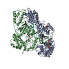
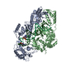
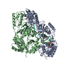
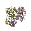
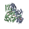
| Title | HIV-1 REVERSE TRANSCRIPTASE COMPLEX WITH DNA AND INHIBITOR RMC-259 | ||||||
 Components Components |
| ||||||
 Keywords Keywords | TRANSFERASE / REVERSE TRANSCRIPTASE / RT INHIBITOR COMPLEX / ACYCLIC NUCLEOSIDE PHOSPHONATE ANALOG / RT-DNA COMPLEX | ||||||
| Function / homology |  Function and homology information Function and homology informationHIV-1 retropepsin / symbiont-mediated activation of host apoptosis / retroviral ribonuclease H / exoribonuclease H / exoribonuclease H activity / DNA integration / viral genome integration into host DNA / establishment of integrated proviral latency / RNA-directed DNA polymerase / RNA stem-loop binding ...HIV-1 retropepsin / symbiont-mediated activation of host apoptosis / retroviral ribonuclease H / exoribonuclease H / exoribonuclease H activity / DNA integration / viral genome integration into host DNA / establishment of integrated proviral latency / RNA-directed DNA polymerase / RNA stem-loop binding / viral penetration into host nucleus / host multivesicular body / RNA-directed DNA polymerase activity / RNA-DNA hybrid ribonuclease activity / Transferases; Transferring phosphorus-containing groups; Nucleotidyltransferases / host cell / viral nucleocapsid / DNA recombination / DNA-directed DNA polymerase / aspartic-type endopeptidase activity / Hydrolases; Acting on ester bonds / DNA-directed DNA polymerase activity / symbiont-mediated suppression of host gene expression / viral translational frameshifting / symbiont entry into host cell / lipid binding / host cell plasma membrane / host cell nucleus / virion membrane / structural molecule activity / proteolysis / DNA binding / zinc ion binding Similarity search - Function | ||||||
| Biological species |  Human immunodeficiency virus type 1 group M subtype BHuman immunodeficiency virus type 1 BH10 Human immunodeficiency virus type 1 group M subtype BHuman immunodeficiency virus type 1 BH10 Homo sapiens (human) Homo sapiens (human) | ||||||
| Method |  X-RAY DIFFRACTION / SYNCHROTRON / MOLECULAR REPLACEMENT / Resolution: 3.1 Å X-RAY DIFFRACTION / SYNCHROTRON / MOLECULAR REPLACEMENT / Resolution: 3.1 Å | ||||||
 Authors Authors | Martinez, S.E. / Singh, A.K. / Gu, W. / Das, K. | ||||||
 Citation Citation | Journal: Eur.J.Med.Chem. / Year: 2021 Title: Exploring the dNTP -binding site of HIV-1 reverse transcriptase for inhibitor design. Authors: Gu, W. / Martinez, S. / Singh, A.K. / Nguyen, H. / Rozenski, J. / Schols, D. / Herdewijn, P. / Das, K. / De Jonghe, S. | ||||||
| History |
|
- Structure visualization
| Structure viewer | Molecule: MolmilJmol/JSmol |
|---|
- Downloads & links
-Download
| PDBx/mmCIF format | 7otz.cif.gz | 456.2 KB | Display | PDBx/mmCIF format |
|---|---|---|---|---|
| PDB format | pdb7otz.ent.gz | 361.6 KB | Display | PDB format |
| PDBx/mmJSON format | 7otz.json.gz | Tree view | PDBx/mmJSON format | |
| Others |  Other downloads Other downloads |
-Validation report
| Arichive directory | https://data.pdbj.org/pub/pdb/validation_reports/ot/7otzftp://data.pdbj.org/pub/pdb/validation_reports/ot/7otz | HTTPS FTP |
|---|
-Related structure data
| Related structure data |  7ot6C  7otaC  7otkC  7otnC  7otxC  7outC 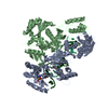 3v4iS C: citing same article ( S: Starting model for refinement |
|---|---|
| Similar structure data |


-Links
 PDBj
PDBj


- Assembly
| Deposited unit | 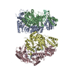
| ||||||||
|---|---|---|---|---|---|---|---|---|---|
| 1 | 
| ||||||||
| 2 | 
| ||||||||
| Unit cell |
|
-Components
-Reverse transcriptase/ribonuclease ... , 2 types, 4 molecules CADB
| #1: Protein | Mass: 64022.414 Da / Num. of mol.: 2 Source method: isolated from a genetically manipulated source Source: (gene. exp.) Human immunodeficiency virus type 1 group M subtype B (isolate BH10)Strain: isolate BH10 / Gene: gag-pol / Production host:  References: UniProt: P03366, RNA-directed DNA polymerase, DNA-directed DNA polymerase, retroviral ribonuclease H, exoribonuclease H #2: Protein | Mass: 50039.488 Da / Num. of mol.: 2 Source method: isolated from a genetically manipulated source Source: (gene. exp.) Human immunodeficiency virus type 1 group M subtype B (isolate BH10)Strain: isolate BH10 / Gene: gag-pol / Production host: References: UniProt: P03366, RNA-directed DNA polymerase, DNA-directed DNA polymerase, retroviral ribonuclease H, exoribonuclease H |
|---|
-DNA chain , 2 types, 4 molecules ETFP
| #3: DNA chain | Mass: 8383.385 Da / Num. of mol.: 2 / Source method: obtained synthetically / Source: (synth.) Human immunodeficiency virus type 1 BH10#4: DNA chain | Mass: 6474.268 Da / Num. of mol.: 2 / Source method: obtained synthetically / Source: (synth.) Homo sapiens (human) |
|---|
-Non-polymers , 2 types, 2 molecules 
| #5: Chemical | ChemComp-MG /  Mass: 24.305 Da / Num. of mol.: 1 / Source method: obtained synthetically / Formula: Mg Mass: 24.305 Da / Num. of mol.: 1 / Source method: obtained synthetically / Formula: Mg |
|---|---|
| #6: Chemical | ChemComp-1IO / ( Mass: 331.222 Da / Num. of mol.: 1 / Source method: obtained synthetically / Formula: C10H14N5O6P / Feature type: SUBJECT OF INVESTIGATION Mass: 331.222 Da / Num. of mol.: 1 / Source method: obtained synthetically / Formula: C10H14N5O6P / Feature type: SUBJECT OF INVESTIGATION |
-Details
| Has ligand of interest | Y |
|---|
-Experimental details
-Experiment
| Experiment | Method: X-RAY DIFFRACTION / Number of used crystals: 1 |
|---|
- Sample preparation
| Crystal | Density Matthews: 3.22 Å3/Da / Density % sol: 61.85 % |
|---|---|
| Crystal grow | Temperature: 277 K / Method: vapor diffusion, sitting drop Details: 16-19% V/V PEG SMEAR BROAD, 0.2 M (NH4)2SO4, 0.1 M TRIS-HCL |
-Data collection
| Diffraction | Mean temperature: 100 K / Serial crystal experiment: N |
|---|---|
| Diffraction source | Source: SYNCHROTRON / Site: SOLEIL  / Beamline: PROXIMA 2 / Wavelength: 0.9801 Å / Beamline: PROXIMA 2 / Wavelength: 0.9801 Å |
| Detector | Type: DECTRIS EIGER X 9M / Detector: PIXEL / Date: Feb 9, 2020 |
| Radiation | Protocol: SINGLE WAVELENGTH / Monochromatic (M) / Laue (L): M / Scattering type: x-ray |
| Radiation wavelength | Wavelength: 0.9801 Å / Relative weight: 1 |
| Reflection | Resolution: 3.1→96.19 Å / Num. obs: 59442 / % possible obs: 100 % / Redundancy: 6.4 % / Biso Wilson estimate: 95.21 Å2 / CC1/2: 0.996 / Rmerge(I) obs: 0.14 / Rpim(I) all: 0.06 / Rrim(I) all: 0.152 / Net I/σ(I): 9.4 / Num. measured all: 381481 / Scaling rejects: 101 |
| Reflection shell | Resolution: 3.1→3.18 Å / Redundancy: 6.5 % / Rmerge(I) obs: 1.84 / Num. unique obs: 4578 / CC1/2: 0.33 / Rpim(I) all: 0.788 / Rrim(I) all: 2.005 / % possible all: 100 |
- Processing
| Software |
| ||||||||||||||||||||||||||||||||||||||||||||||||||||||||||||||||||||||||||||||||||||
|---|---|---|---|---|---|---|---|---|---|---|---|---|---|---|---|---|---|---|---|---|---|---|---|---|---|---|---|---|---|---|---|---|---|---|---|---|---|---|---|---|---|---|---|---|---|---|---|---|---|---|---|---|---|---|---|---|---|---|---|---|---|---|---|---|---|---|---|---|---|---|---|---|---|---|---|---|---|---|---|---|---|---|---|---|---|
| Refinement | Method to determine structure: MOLECULAR REPLACEMENT Starting model: 3V4I Resolution: 3.1→96.19 Å / SU ML: 0.49 / Cross valid method: THROUGHOUT / σ(F): 1.34 / Phase error: 29.75 / Stereochemistry target values: ML
| ||||||||||||||||||||||||||||||||||||||||||||||||||||||||||||||||||||||||||||||||||||
| Solvent computation | Shrinkage radii: 0.9 Å / VDW probe radii: 1.11 Å / Solvent model: FLAT BULK SOLVENT MODEL | ||||||||||||||||||||||||||||||||||||||||||||||||||||||||||||||||||||||||||||||||||||
| Displacement parameters | Biso max: 299.26 Å2 / Biso mean: 115.0933 Å2 / Biso min: 28.26 Å2 | ||||||||||||||||||||||||||||||||||||||||||||||||||||||||||||||||||||||||||||||||||||
| Refinement step | Cycle: final / Resolution: 3.1→96.19 Å
| ||||||||||||||||||||||||||||||||||||||||||||||||||||||||||||||||||||||||||||||||||||
| LS refinement shell | Refine-ID: X-RAY DIFFRACTION / Rfactor Rfree error: 0 / Total num. of bins used: 13 / % reflection obs: 100 %
|
