 Movie
Movie Controller


+ Open data

- Basic information
| Entry | Database: PDB / ID: 7ose | ||||||
|---|---|---|---|---|---|---|---|
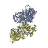
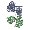
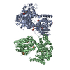
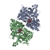
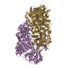
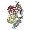
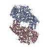
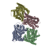
| Title | cytochrome bd-II type oxidase with bound aurachin D | ||||||
 Components Components |
| ||||||
 Keywords Keywords | MEMBRANE PROTEIN / terminal oxidase / Q-loop / inhibitor binding | ||||||
| Function / homology |  Function and homology information Function and homology informationubiquinol oxidase (H+-transporting) / cytochrome bo3 ubiquinol oxidase activity / cytochrome complex / aerobic electron transport chain / oxidoreductase activity, acting on diphenols and related substances as donors, oxygen as acceptor / cell outer membrane / electron transfer activity / heme binding / membrane / metal ion binding / plasma membrane Similarity search - Function | ||||||
| Biological species |  | ||||||
| Method | ELECTRON MICROSCOPY / single particle reconstruction / cryo EM / Resolution: 3 Å | ||||||
 Authors Authors | Grauel, A. / Kaegi, J. / Rasmussen, T. / Wohlwend, D. / Boettcher, B. / Friedrich, T. | ||||||
| Funding support |  Germany, 1items Germany, 1items
| ||||||
 Citation Citation | Journal: Nat Commun / Year: 2021 Title: Structure of Escherichia coli cytochrome bd-II type oxidase with bound aurachin D. Authors: Antonia Grauel / Jan Kägi / Tim Rasmussen / Iryna Makarchuk / Sabrina Oppermann / Aurélien F A Moumbock / Daniel Wohlwend / Rolf Müller / Frederic Melin / Stefan Günther / Petra Hellwig ...Authors: Antonia Grauel / Jan Kägi / Tim Rasmussen / Iryna Makarchuk / Sabrina Oppermann / Aurélien F A Moumbock / Daniel Wohlwend / Rolf Müller / Frederic Melin / Stefan Günther / Petra Hellwig / Bettina Böttcher / Thorsten Friedrich /  Abstract: Cytochrome bd quinol:O oxidoreductases are respiratory terminal oxidases so far only identified in prokaryotes, including several pathogenic bacteria. Escherichia coli contains two bd oxidases of ...Cytochrome bd quinol:O oxidoreductases are respiratory terminal oxidases so far only identified in prokaryotes, including several pathogenic bacteria. Escherichia coli contains two bd oxidases of which only the bd-I type is structurally characterized. Here, we report the structure of the Escherichia coli cytochrome bd-II type oxidase with the bound inhibitor aurachin D as obtained by electron cryo-microscopy at 3 Å resolution. The oxidase consists of subunits AppB, C and X that show an architecture similar to that of bd-I. The three heme cofactors are found in AppC, while AppB is stabilized by a structural ubiquinone-8 at the homologous positions. A fourth subunit present in bd-I is lacking in bd-II. Accordingly, heme b is exposed to the membrane but heme d embedded within the protein and showing an unexpectedly high redox potential is the catalytically active centre. The structure of the Q-loop is fully resolved, revealing the specific aurachin binding. | ||||||
| History |
|
- Structure visualization
| Movie |
Movie viewer |
|---|---|
| Structure viewer | Molecule: MolmilJmol/JSmol |
- Downloads & links
-Download
| PDBx/mmCIF format | 7ose.cif.gz | 333.8 KB | Display | PDBx/mmCIF format |
|---|---|---|---|---|
| PDB format | pdb7ose.ent.gz | 269.4 KB | Display | PDB format |
| PDBx/mmJSON format | 7ose.json.gz | Tree view | PDBx/mmJSON format | |
| Others |  Other downloads Other downloads |
-Validation report
| Arichive directory | https://data.pdbj.org/pub/pdb/validation_reports/os/7oseftp://data.pdbj.org/pub/pdb/validation_reports/os/7ose | HTTPS FTP |
|---|
-Related structure data
| Related structure data |  13048MC M: map data used to model this data C: citing same article ( |
|---|---|
| Similar structure data |


-Links
 PDBj
PDBj
- Assembly
| Deposited unit | 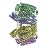
|
|---|---|
| 1 |
|
-Components
-Cytochrome bd-II ubiquinol oxidase subunit ... , 2 types, 4 molecules ADBE
| #1: Protein | Mass: 57962.469 Da / Num. of mol.: 2 Source method: isolated from a genetically manipulated source Source: (gene. exp.) References: UniProt: P26459, ubiquinol oxidase (H+-transporting) #2: Protein | Mass: 42448.543 Da / Num. of mol.: 2 Source method: isolated from a genetically manipulated source Source: (gene. exp.) References: UniProt: P26458, ubiquinol oxidase (H+-transporting) |
|---|
-Protein/peptide , 1 types, 2 molecules CF
| #3: Protein/peptide | Mass: 3599.463 Da / Num. of mol.: 2 Source method: isolated from a genetically manipulated source Source: (gene. exp.) |
|---|
-Non-polymers , 5 types, 12 molecules 


| #4: Chemical | ChemComp-HEB /  Mass: 618.503 Da / Num. of mol.: 4 / Source method: obtained synthetically / Formula: C34H34FeN4O4 Mass: 618.503 Da / Num. of mol.: 4 / Source method: obtained synthetically / Formula: C34H34FeN4O4#5: Chemical |  Mass: 632.487 Da / Num. of mol.: 2 / Source method: obtained synthetically / Formula: C34H32FeN4O5 Mass: 632.487 Da / Num. of mol.: 2 / Source method: obtained synthetically / Formula: C34H32FeN4O5#6: Chemical |  Mass: 363.536 Da / Num. of mol.: 2 / Source method: obtained synthetically / Formula: C25H33NO Mass: 363.536 Da / Num. of mol.: 2 / Source method: obtained synthetically / Formula: C25H33NO#7: Chemical |  Mass: 727.109 Da / Num. of mol.: 2 / Source method: obtained synthetically / Formula: C49H74O4 Mass: 727.109 Da / Num. of mol.: 2 / Source method: obtained synthetically / Formula: C49H74O4#8: Water | ChemComp-HOH / | Mass: 18.015 Da / Num. of mol.: 2 / Source method: isolated from a natural source / Formula: H2O |
|---|
-Details
| Has ligand of interest | N |
|---|
-Experimental details
-Experiment
| Experiment | Method: ELECTRON MICROSCOPY |
|---|---|
| EM experiment | Aggregation state: PARTICLE / 3D reconstruction method: single particle reconstruction |
- Sample preparation
| Component | Name: Cytochrome bd-II ubiquinol oxidase / Type: COMPLEX Details: dimer of heterotrimers solubilised in amphipol A8-35 in complex with the inhibitor Aurachin D Entity ID: #1-#3 / Source: RECOMBINANT | ||||||||||||||||||||
|---|---|---|---|---|---|---|---|---|---|---|---|---|---|---|---|---|---|---|---|---|---|
| Molecular weight | Value: 0.208 MDa / Experimental value: NO | ||||||||||||||||||||
| Source (natural) | Organism: | ||||||||||||||||||||
| Source (recombinant) | Organism: | ||||||||||||||||||||
| Buffer solution | pH: 7 | ||||||||||||||||||||
| Buffer component |
| ||||||||||||||||||||
| Specimen | Conc.: 5.6 mg/ml / Embedding applied: NO / Shadowing applied: NO / Staining applied: NO / Vitrification applied: YES | ||||||||||||||||||||
| Specimen support | Grid material: COPPER / Grid mesh size: 400 divisions/in. / Grid type: Quantifoil R1.2/1.3 | ||||||||||||||||||||
| Vitrification | Instrument: FEI VITROBOT MARK IV / Cryogen name: ETHANE / Humidity: 100 % / Chamber temperature: 277 K / Details: 45 sec incubation, 6.5 sec blotting |
- Electron microscopy imaging
| Experimental equipment |  Model: Titan Krios / Image courtesy: FEI Company |
|---|---|
| Microscopy | Model: FEI TITAN KRIOS |
| Electron gun | Electron source:  FIELD EMISSION GUN / Accelerating voltage: 300 kV / Illumination mode: FLOOD BEAM FIELD EMISSION GUN / Accelerating voltage: 300 kV / Illumination mode: FLOOD BEAM |
| Electron lens | Mode: BRIGHT FIELD / Nominal magnification: 75000 X / Nominal defocus max: 2400 nm / Nominal defocus min: 1400 nm / Cs: 2.7 mm / C2 aperture diameter: 70 µm / Alignment procedure: COMA FREE |
| Specimen holder | Cryogen: NITROGEN / Specimen holder model: FEI TITAN KRIOS AUTOGRID HOLDER |
| Image recording | Average exposure time: 75 sec. / Electron dose: 79 e/Å2 / Detector mode: COUNTING / Film or detector model: FEI FALCON III (4k x 4k) / Num. of grids imaged: 1 / Num. of real images: 1836 |
- Processing
| Software | Name: PHENIX / Version: 1.19_4092: / Classification: refinement | ||||||||||||||||||||||||||||||||||||||||
|---|---|---|---|---|---|---|---|---|---|---|---|---|---|---|---|---|---|---|---|---|---|---|---|---|---|---|---|---|---|---|---|---|---|---|---|---|---|---|---|---|---|
| EM software |
| ||||||||||||||||||||||||||||||||||||||||
| CTF correction | Type: PHASE FLIPPING AND AMPLITUDE CORRECTION | ||||||||||||||||||||||||||||||||||||||||
| Particle selection | Num. of particles selected: 800000 | ||||||||||||||||||||||||||||||||||||||||
| Symmetry | Point symmetry: C2 (2 fold cyclic) | ||||||||||||||||||||||||||||||||||||||||
| 3D reconstruction | Resolution: 3 Å / Resolution method: FSC 0.143 CUT-OFF / Num. of particles: 125497 / Algorithm: FOURIER SPACE / Symmetry type: POINT | ||||||||||||||||||||||||||||||||||||||||
| Atomic model building | B value: 94 / Protocol: FLEXIBLE FIT / Space: REAL / Target criteria: WEIGHTED MAP SUM AT ATOM CENTERS | ||||||||||||||||||||||||||||||||||||||||
| Atomic model building | PDB-ID: 6RX4 Accession code: 6RX4 / Source name: PDB / Type: experimental model | ||||||||||||||||||||||||||||||||||||||||
| Refine LS restraints |
|

