 Movie
Movie Controller


+ Open data

- Basic information
| Entry | Database: EMDB / ID: EMD-13048 | |||||||||
|---|---|---|---|---|---|---|---|---|---|---|
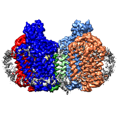
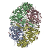
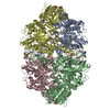
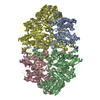
| Title | cytochrome bd-II type oxidase with bound aurachin D | |||||||||
 Map data Map data | post-processed sharpened map | |||||||||
 Sample Sample |
| |||||||||
 Keywords Keywords | terminal oxidase / Q-loop / inhibitor binding / MEMBRANE PROTEIN | |||||||||
| Function / homology |  Function and homology information Function and homology informationubiquinol oxidase (H+-transporting) / cytochrome bo3 ubiquinol oxidase activity / cytochrome complex / aerobic electron transport chain / oxidoreductase activity, acting on diphenols and related substances as donors, oxygen as acceptor / cell outer membrane / electron transfer activity / heme binding / membrane / metal ion binding / plasma membrane Similarity search - Function | |||||||||
| Biological species |  | |||||||||
| Method | single particle reconstruction / cryo EM / Resolution: 3.0 Å | |||||||||
 Authors Authors | Grauel A / Kaegi J | |||||||||
| Funding support |  Germany, 1 items Germany, 1 items
| |||||||||
 Citation Citation | Journal: Nat Commun / Year: 2021 Title: Structure of Escherichia coli cytochrome bd-II type oxidase with bound aurachin D. Authors: Antonia Grauel / Jan Kägi / Tim Rasmussen / Iryna Makarchuk / Sabrina Oppermann / Aurélien F A Moumbock / Daniel Wohlwend / Rolf Müller / Frederic Melin / Stefan Günther / Petra Hellwig ...Authors: Antonia Grauel / Jan Kägi / Tim Rasmussen / Iryna Makarchuk / Sabrina Oppermann / Aurélien F A Moumbock / Daniel Wohlwend / Rolf Müller / Frederic Melin / Stefan Günther / Petra Hellwig / Bettina Böttcher / Thorsten Friedrich /  Abstract: Cytochrome bd quinol:O oxidoreductases are respiratory terminal oxidases so far only identified in prokaryotes, including several pathogenic bacteria. Escherichia coli contains two bd oxidases of ...Cytochrome bd quinol:O oxidoreductases are respiratory terminal oxidases so far only identified in prokaryotes, including several pathogenic bacteria. Escherichia coli contains two bd oxidases of which only the bd-I type is structurally characterized. Here, we report the structure of the Escherichia coli cytochrome bd-II type oxidase with the bound inhibitor aurachin D as obtained by electron cryo-microscopy at 3 Å resolution. The oxidase consists of subunits AppB, C and X that show an architecture similar to that of bd-I. The three heme cofactors are found in AppC, while AppB is stabilized by a structural ubiquinone-8 at the homologous positions. A fourth subunit present in bd-I is lacking in bd-II. Accordingly, heme b is exposed to the membrane but heme d embedded within the protein and showing an unexpectedly high redox potential is the catalytically active centre. The structure of the Q-loop is fully resolved, revealing the specific aurachin binding. | |||||||||
| History |
|
- Structure visualization
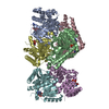
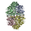
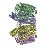
| Movie |
Movie viewer |
|---|---|
| Structure viewer | EM map: SurfViewMolmilJmol/JSmol |
| Supplemental images |
- Downloads & links
-EMDB archive
| Map data | emd_13048.map.gz | 27.2 MB | EMDB map data format | |
|---|---|---|---|---|
| Header (meta data) | emd-13048-v30.xmlemd-13048.xml | 19 KB 19 KB | Display Display | EMDB header |
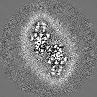
| Images |  emd_13048.png emd_13048.png | 167.6 KB | ||
| Archive directory |  http://ftp.pdbj.org/pub/emdb/structures/EMD-13048ftp://ftp.pdbj.org/pub/emdb/structures/EMD-13048 http://ftp.pdbj.org/pub/emdb/structures/EMD-13048ftp://ftp.pdbj.org/pub/emdb/structures/EMD-13048 | HTTPS FTP |
-Related structure data
| Related structure data |  7oseMC M: atomic model generated by this map C: citing same article ( |
|---|---|
| Similar structure data |


-Links
| EMDB pages | EMDB (EBI/PDBe) / EMDataResource |
|---|
-Map
| File | Download / File: emd_13048.map.gz / Format: CCP4 / Size: 30.5 MB / Type: IMAGE STORED AS FLOATING POINT NUMBER (4 BYTES) | ||||||||||||||||||||||||||||||||||||||||||||||||||||||||||||
|---|---|---|---|---|---|---|---|---|---|---|---|---|---|---|---|---|---|---|---|---|---|---|---|---|---|---|---|---|---|---|---|---|---|---|---|---|---|---|---|---|---|---|---|---|---|---|---|---|---|---|---|---|---|---|---|---|---|---|---|---|---|
| Annotation | post-processed sharpened map | ||||||||||||||||||||||||||||||||||||||||||||||||||||||||||||
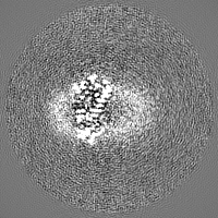
| Projections & slices | Image control
Images are generated by Spider. | ||||||||||||||||||||||||||||||||||||||||||||||||||||||||||||
| Voxel size | X=Y=Z: 1.0635 Å | ||||||||||||||||||||||||||||||||||||||||||||||||||||||||||||
| Density |
| ||||||||||||||||||||||||||||||||||||||||||||||||||||||||||||
| Symmetry | Space group: 1 | ||||||||||||||||||||||||||||||||||||||||||||||||||||||||||||
| Details | EMDB XML:
CCP4 map header:
| ||||||||||||||||||||||||||||||||||||||||||||||||||||||||||||
 Z (Sec.)
 Y (Row.)
 X (Col.)


-Supplemental data
- Sample components
-Entire : Cytochrome bd-II ubiquinol oxidase
| Entire | Name: Cytochrome bd-II ubiquinol oxidase |
|---|---|
| Components |
|
-Supramolecule #1: Cytochrome bd-II ubiquinol oxidase
| Supramolecule | Name: Cytochrome bd-II ubiquinol oxidase / type: complex / ID: 1 / Parent: 0 / Macromolecule list: #1-#3 Details: dimer of heterotrimers solubilised in amphipol A8-35 in complex with the inhibitor Aurachin D |
|---|---|
| Source (natural) | Organism: |
| Molecular weight | Theoretical: 208 KDa |
-Macromolecule #1: Cytochrome bd-II ubiquinol oxidase subunit 1
| Macromolecule | Name: Cytochrome bd-II ubiquinol oxidase subunit 1 / type: protein_or_peptide / ID: 1 / Number of copies: 2 / Enantiomer: LEVO / EC number: ubiquinol oxidase (H+-transporting) |
|---|---|
| Source (natural) | Organism: |
| Molecular weight | Theoretical: 57.962469 KDa |
| Recombinant expression | Organism: |
| Sequence | String: MWDVIDLSRW QFALTALYHF LFVPLTLGLI FLLAIMETIY VVTGKTIYRD MTRFWGKLFG INFALGVATG LTMEFQFGTN WSFYSNYVG DIFGAPLAME ALMAFFLEST FVGLFFFGWQ RLNKYQHLLV TWLVAFGSNL SALWILNANG WMQYPTGAHF D IDTLRMEM ...String: MWDVIDLSRW QFALTALYHF LFVPLTLGLI FLLAIMETIY VVTGKTIYRD MTRFWGKLFG INFALGVATG LTMEFQFGTN WSFYSNYVG DIFGAPLAME ALMAFFLEST FVGLFFFGWQ RLNKYQHLLV TWLVAFGSNL SALWILNANG WMQYPTGAHF D IDTLRMEM TSFSELVFNP VSQVKFVHTV MAGYVTGAMF IMAISAWYLL RGRERNVALR SFAIGSVFGT LAIIGTLQLG DS SAYEVAQ VQPVKLAAME GEWQTEPAPA PFHVVAWPEQ DQERNAFALK IPALLGILAT HSLDKPVPGL KNLMAETYPR LQR GRMAWL LMQEISQGNR EPHVLQAFRG LEGDLGYGML LSRYAPDMNH VTAAQYQAAM RGAIPQVAPV FWSFRIMVGC GSLL LLVML IALVQTLRGK IDQHRWVLKM ALWSLPLPWI AIEAGWFMTE FGRQPWAIQD ILPTYSAHSA LTTGQLAFSL IMIVG LYTL FLIAEVYLMQ KYARLGPSAM QSEQPTQQQG UniProtKB: Cytochrome bd-II ubiquinol oxidase subunit 1 |
-Macromolecule #2: Cytochrome bd-II ubiquinol oxidase subunit 2
| Macromolecule | Name: Cytochrome bd-II ubiquinol oxidase subunit 2 / type: protein_or_peptide / ID: 2 / Number of copies: 2 / Enantiomer: LEVO / EC number: ubiquinol oxidase (H+-transporting) |
|---|---|
| Source (natural) | Organism: |
| Molecular weight | Theoretical: 42.448543 KDa |
| Recombinant expression | Organism: |
| Sequence | String: MFDYETLRFI WWLLIGVILV VFMISDGFDM GIGCLLPLVA RNDDERRIVI NSVGAHWEGN QVWLILAGGA LFAAWPRVYA AAFSGFYVA MILVLCSLFF RPLAFDYRGK IADARWRKMW DAGLVIGSLV PPVVFGIAFG NLLLGVPFAF TPQLRVEYLG S FWQLLTPF ...String: MFDYETLRFI WWLLIGVILV VFMISDGFDM GIGCLLPLVA RNDDERRIVI NSVGAHWEGN QVWLILAGGA LFAAWPRVYA AAFSGFYVA MILVLCSLFF RPLAFDYRGK IADARWRKMW DAGLVIGSLV PPVVFGIAFG NLLLGVPFAF TPQLRVEYLG S FWQLLTPF PLLCGLLSLG MVILQGGVWL QLKTVGVIHL RSQLATKRAA LLVMLCFLLA GYWLWVGIDG FVLLAQDANG PS NPLMKLV AVLPGAWMNN FVESPVLWIF PLLGFFCPLL TVMAIYRGRP GWGFLMASLM QFGVIFTAGI TLFPFVMPSS VSP ISSLTL WDSTSSQLTL SIMLVIVLIF LPIVLLYTLW SYYKMWGRMT TETLRRNENE LY UniProtKB: Cytochrome bd-II ubiquinol oxidase subunit 2 |
-Macromolecule #3: Putative cytochrome bd-II ubiquinol oxidase subunit AppX
| Macromolecule | Name: Putative cytochrome bd-II ubiquinol oxidase subunit AppX type: protein_or_peptide / ID: 3 / Number of copies: 2 / Enantiomer: LEVO |
|---|---|
| Source (natural) | Organism: |
| Molecular weight | Theoretical: 3.599463 KDa |
| Recombinant expression | Organism: |
| Sequence | String: MWYLLWFVGI LLMCSLSTLV LVWLDPRLKS UniProtKB: Putative cytochrome bd-II ubiquinol oxidase subunit AppX |
-Macromolecule #4: HEME B/C
| Macromolecule | Name: HEME B/C / type: ligand / ID: 4 / Number of copies: 4 / Formula: HEB |
|---|---|
| Molecular weight | Theoretical: 618.503 Da |
| Chemical component information |  ChemComp-HEB: |
-Macromolecule #5: CIS-HEME D HYDROXYCHLORIN GAMMA-SPIROLACTONE
| Macromolecule | Name: CIS-HEME D HYDROXYCHLORIN GAMMA-SPIROLACTONE / type: ligand / ID: 5 / Number of copies: 2 / Formula: HDD |
|---|---|
| Molecular weight | Theoretical: 632.487 Da |
| Chemical component information |  ChemComp-HDD: |
-Macromolecule #6: Aurachin D
| Macromolecule | Name: Aurachin D / type: ligand / ID: 6 / Number of copies: 2 / Formula: 0NI |
|---|---|
| Molecular weight | Theoretical: 363.536 Da |
| Chemical component information |  ChemComp-0NI: |
-Macromolecule #7: Ubiquinone-8
| Macromolecule | Name: Ubiquinone-8 / type: ligand / ID: 7 / Number of copies: 2 / Formula: UQ8 |
|---|---|
| Molecular weight | Theoretical: 727.109 Da |
| Chemical component information |  ChemComp-UQ8: |
-Macromolecule #8: water
| Macromolecule | Name: water / type: ligand / ID: 8 / Number of copies: 2 / Formula: HOH |
|---|---|
| Molecular weight | Theoretical: 18.015 Da |
| Chemical component information |  ChemComp-HOH: |
-Experimental details
-Structure determination
| Method | cryo EM |
|---|---|
 Processing Processing | single particle reconstruction |
| Aggregation state | particle |
-Sample preparation
| Concentration | 5.6 mg/mL | ||||||||||||
|---|---|---|---|---|---|---|---|---|---|---|---|---|---|
| Buffer | pH: 7 Component:
| ||||||||||||
| Grid | Model: Quantifoil R1.2/1.3 / Material: COPPER / Mesh: 400 / Support film - Material: CARBON / Support film - topology: HOLEY ARRAY / Pretreatment - Type: GLOW DISCHARGE / Pretreatment - Time: 150 sec. / Pretreatment - Atmosphere: AIR | ||||||||||||
| Vitrification | Cryogen name: ETHANE / Chamber humidity: 100 % / Chamber temperature: 277 K / Instrument: FEI VITROBOT MARK IV / Details: 45 sec incubation, 6.5 sec blotting. |
- Electron microscopy
| Microscope | FEI TITAN KRIOS |
|---|---|
| Image recording | Film or detector model: FEI FALCON III (4k x 4k) / Detector mode: COUNTING / Number grids imaged: 1 / Number real images: 1836 / Average exposure time: 75.0 sec. / Average electron dose: 79.0 e/Å2 |
| Electron beam | Acceleration voltage: 300 kV / Electron source:  FIELD EMISSION GUN FIELD EMISSION GUN |
| Electron optics | C2 aperture diameter: 70.0 µm / Illumination mode: FLOOD BEAM / Imaging mode: BRIGHT FIELD / Cs: 2.7 mm / Nominal defocus max: 2.4 µm / Nominal defocus min: 1.4000000000000001 µm / Nominal magnification: 75000 |
| Sample stage | Specimen holder model: FEI TITAN KRIOS AUTOGRID HOLDER / Cooling holder cryogen: NITROGEN |
| Experimental equipment |  Model: Titan Krios / Image courtesy: FEI Company |

