 Movie
Movie Controller


+ Open data

- Basic information
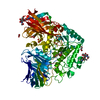
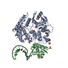
| Entry | Database: PDB / ID: 6rx4 | |||||||||
|---|---|---|---|---|---|---|---|---|---|---|
| Title | THE STRUCTURE OF BD OXIDASE FROM ESCHERICHIA COLI | |||||||||
 Components Components | (Cytochrome bd-I ubiquinol oxidase subunit ...) x 4 | |||||||||
 Keywords Keywords | OXIDOREDUCTASE / BD OXIDASE / TERMINAL OXIDASE | |||||||||
| Function / homology |  Function and homology information Function and homology informationquinol oxidase (electrogenic, proton-motive force generating) / oxidoreductase activity, acting on diphenols and related substances as donors / cytochrome complex / aerobic electron transport chain / outer membrane / oxidoreductase activity, acting on diphenols and related substances as donors, oxygen as acceptor / oxidative phosphorylation / electron transfer activity / heme binding / membrane ...quinol oxidase (electrogenic, proton-motive force generating) / oxidoreductase activity, acting on diphenols and related substances as donors / cytochrome complex / aerobic electron transport chain / outer membrane / oxidoreductase activity, acting on diphenols and related substances as donors, oxygen as acceptor / oxidative phosphorylation / electron transfer activity / heme binding / membrane / metal ion binding / plasma membrane Similarity search - Function | |||||||||
| Biological species |  | |||||||||
| Method | ELECTRON MICROSCOPY / single particle reconstruction / cryo EM / Resolution: 3.3 Å | |||||||||
 Authors Authors | Rasmussen, T. / Boettcher, B. / Thesseling, A. / Friedrich, T. | |||||||||
| Funding support |  Germany, 1items Germany, 1items
| |||||||||
 Citation Citation | Journal: Nat Commun / Year: 2019 Title: Homologous bd oxidases share the same architecture but differ in mechanism. Authors: Alexander Theßeling / Tim Rasmussen / Sabrina Burschel / Daniel Wohlwend / Jan Kägi / Rolf Müller / Bettina Böttcher / Thorsten Friedrich / Abstract: Cytochrome bd oxidases are terminal reductases of bacterial and archaeal respiratory chains. The enzyme couples the oxidation of ubiquinol or menaquinol with the reduction of dioxygen to water, thus ...Cytochrome bd oxidases are terminal reductases of bacterial and archaeal respiratory chains. The enzyme couples the oxidation of ubiquinol or menaquinol with the reduction of dioxygen to water, thus contributing to the generation of the protonmotive force. Here, we determine the structure of the Escherichia coli bd oxidase treated with the specific inhibitor aurachin by cryo-electron microscopy (cryo-EM). The major subunits CydA and CydB are related by a pseudo two fold symmetry. The heme b and d cofactors are found in CydA, while ubiquinone-8 is bound at the homologous positions in CydB to stabilize its structure. The architecture of the E. coli enzyme is highly similar to that of Geobacillus thermodenitrificans, however, the positions of heme b and d are interchanged, and a common oxygen channel is blocked by a fourth subunit and substituted by a more narrow, alternative channel. Thus, with the same overall fold, the homologous enzymes exhibit a different mechanism. | |||||||||
| History |
|
- Structure visualization
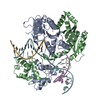
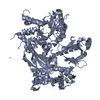
| Movie |
Movie viewer |
|---|---|
| Structure viewer | Molecule: MolmilJmol/JSmol |
- Downloads & links
-Download
| PDBx/mmCIF format | 6rx4.cif.gz | 177.6 KB | Display | PDBx/mmCIF format |
|---|---|---|---|---|
| PDB format | pdb6rx4.ent.gz | 136.4 KB | Display | PDB format |
| PDBx/mmJSON format | 6rx4.json.gz | Tree view | PDBx/mmJSON format | |
| Others |  Other downloads Other downloads |
-Validation report
| Arichive directory | https://data.pdbj.org/pub/pdb/validation_reports/rx/6rx4ftp://data.pdbj.org/pub/pdb/validation_reports/rx/6rx4 | HTTPS FTP |
|---|
-Related structure data
| Related structure data |  10049MC M: map data used to model this data C: citing same article ( |
|---|---|
| Similar structure data |


-Links
 PDBj
PDBj

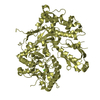
- Assembly
| Deposited unit | 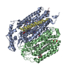
|
|---|---|
| 1 |
|
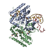
-Components
-Cytochrome bd-I ubiquinol oxidase subunit ... , 4 types, 4 molecules ABCD
| #1: Protein | Mass: 58251.723 Da / Num. of mol.: 1 / Source method: isolated from a natural source / Source: (natural) References: UniProt: P0ABJ9, quinol oxidase (electrogenic, proton-motive force generating) |
|---|---|
| #2: Protein | Mass: 42479.828 Da / Num. of mol.: 1 / Source method: isolated from a natural source / Source: (natural) References: UniProt: P0ABK2, quinol oxidase (electrogenic, proton-motive force generating) |
| #3: Protein/peptide | Mass: 4043.663 Da / Num. of mol.: 1 / Source method: isolated from a natural source / Source: (natural) References: UniProt: P56100, quinol oxidase (electrogenic, proton-motive force generating) |
| #4: Protein/peptide | Mass: 2230.741 Da / Num. of mol.: 1 / Source method: isolated from a natural source / Source: (natural) |
-Non-polymers , 5 types, 12 molecules 


| #5: Chemical |  Mass: 618.503 Da / Num. of mol.: 2 / Source method: obtained synthetically / Formula: C34H34FeN4O4 Mass: 618.503 Da / Num. of mol.: 2 / Source method: obtained synthetically / Formula: C34H34FeN4O4#6: Chemical | ChemComp-HDD / |  Mass: 632.487 Da / Num. of mol.: 1 / Source method: obtained synthetically / Formula: C34H32FeN4O5 Mass: 632.487 Da / Num. of mol.: 1 / Source method: obtained synthetically / Formula: C34H32FeN4O5#7: Chemical | ChemComp-PEE / |  Mass: 744.034 Da / Num. of mol.: 1 / Source method: obtained synthetically / Formula: C41H78NO8P / Comment: DOPE, phospholipid*YM Mass: 744.034 Da / Num. of mol.: 1 / Source method: obtained synthetically / Formula: C41H78NO8P / Comment: DOPE, phospholipid*YM#8: Chemical | ChemComp-UQ8 / |  Mass: 727.109 Da / Num. of mol.: 1 / Source method: obtained synthetically / Formula: C49H74O4 Mass: 727.109 Da / Num. of mol.: 1 / Source method: obtained synthetically / Formula: C49H74O4#9: Water | ChemComp-HOH / | Mass: 18.015 Da / Num. of mol.: 7 / Source method: isolated from a natural source / Formula: H2O |
|---|
-Experimental details
-Experiment
| Experiment | Method: ELECTRON MICROSCOPY |
|---|---|
| EM experiment | Aggregation state: PARTICLE / 3D reconstruction method: single particle reconstruction |
- Sample preparation
| Component | Name: bd-oxidase from Escherichia coli / Type: COMPLEX Details: Reconstruction into Amphipole A8-35 in the presence of the inhibitor aurachin C. Entity ID: #1-#4 / Source: NATURAL | |||||||||||||||
|---|---|---|---|---|---|---|---|---|---|---|---|---|---|---|---|---|
| Source (natural) | Organism: | |||||||||||||||
| Buffer solution | pH: 7.5 | |||||||||||||||
| Buffer component |
| |||||||||||||||
| Specimen | Conc.: 2 mg/ml / Embedding applied: NO / Shadowing applied: NO / Staining applied: NO / Vitrification applied: YES | |||||||||||||||
| Specimen support | Grid material: COPPER / Grid mesh size: 400 divisions/in. / Grid type: Quantifoil R1.2/1.3 | |||||||||||||||
| Vitrification | Instrument: FEI VITROBOT MARK IV / Cryogen name: ETHANE / Humidity: 100 % / Chamber temperature: 277 K / Details: blot time 3.5 sec, blot force 5 |
- Electron microscopy imaging
| Experimental equipment |  Model: Titan Krios / Image courtesy: FEI Company |
|---|---|
| Microscopy | Model: FEI TITAN KRIOS |
| Electron gun | Electron source:  FIELD EMISSION GUN / Accelerating voltage: 300 kV / Illumination mode: FLOOD BEAM FIELD EMISSION GUN / Accelerating voltage: 300 kV / Illumination mode: FLOOD BEAM |
| Electron lens | Mode: BRIGHT FIELD / Nominal magnification: 75000 X / Nominal defocus max: 2200 nm / Nominal defocus min: 1400 nm / Cs: 2.7 mm / C2 aperture diameter: 70 µm / Alignment procedure: COMA FREE |
| Specimen holder | Cryogen: NITROGEN / Specimen holder model: FEI TITAN KRIOS AUTOGRID HOLDER |
| Image recording | Average exposure time: 75 sec. / Electron dose: 59 e/Å2 / Detector mode: COUNTING / Film or detector model: FEI FALCON III (4k x 4k) / Num. of grids imaged: 1 / Num. of real images: 8663 Details: Images were collected in movie mode with 47 frames. |
- Processing
| EM software |
| ||||||||||||||||||||||||||||||||||||||||
|---|---|---|---|---|---|---|---|---|---|---|---|---|---|---|---|---|---|---|---|---|---|---|---|---|---|---|---|---|---|---|---|---|---|---|---|---|---|---|---|---|---|
| Image processing | Details: Movies were motion corrected and dose weighted with the program Motioncorr2. | ||||||||||||||||||||||||||||||||||||||||
| CTF correction | Type: PHASE FLIPPING AND AMPLITUDE CORRECTION | ||||||||||||||||||||||||||||||||||||||||
| Symmetry | Point symmetry: C1 (asymmetric) | ||||||||||||||||||||||||||||||||||||||||
| 3D reconstruction | Resolution: 3.3 Å / Resolution method: FSC 0.143 CUT-OFF / Num. of particles: 197805 / Algorithm: FOURIER SPACE / Symmetry type: POINT | ||||||||||||||||||||||||||||||||||||||||
| Atomic model building | B value: 88 / Protocol: AB INITIO MODEL / Space: REAL | ||||||||||||||||||||||||||||||||||||||||
| Atomic model building | PDB-ID: 5DOQ Accession code: 5DOQ / Source name: PDB / Type: experimental model |

