- PDB-5doq: The structure of bd oxidase from Geobacillus thermodenitrificans -
+
Open data
ID or keywords:
Loading...
-
Basic information
Entry
Database: PDB / ID: 5doq
Title
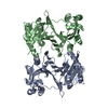
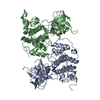
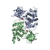
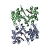
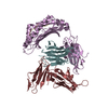
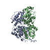
The structure of bd oxidase from Geobacillus thermodenitrificans
Components
Bd-type quinol oxidase subunit I
Bd-type quinol oxidase subunit II
Putative membrane protein
Keywords
OXIDOREDUCTASE / bd oxidase / terminal oxidase
Function / homology
Function and homology information
cytochrome complex / aerobic electron transport chain / oxidoreductase activity, acting on diphenols and related substances as donors, oxygen as acceptor / electron transfer activity / heme binding / membrane / metal ion binding / plasma membrane Similarity search - Function
Protocol: SINGLE WAVELENGTH / Monochromatic (M) / Laue (L): M / Scattering type: x-ray
Radiation wavelength
Wavelength: 1.73829 Å / Relative weight: 1
Reflection
Resolution: 3.05→50 Å / Num. obs: 18373 / % possible obs: 56.7 % / Redundancy: 31.44 % / Rmerge(I) obs: 0.099 / Rsym value: 0.097 / Net I/σ(I): 19.17
Reflection shell
Resolution: 3.05→3.12 Å / Redundancy: 19.15 % / Rmerge(I) obs: 2.2 / Mean I/σ(I) obs: 3.03 / % possible all: 2.6
-
Processing
Software
Name
Version
Classification
REFMAC
5.8.0073
refinement
XDS
datareduction
XSCALE
datascaling
SHELX
phasing
PHENIX
phasing
Refinement
Method to determine structure: SAD / Resolution: 3.05→20 Å / Cor.coef. Fo:Fc: 0.879 / Cor.coef. Fo:Fc free: 0.873 / SU B: 36.631 / SU ML: 0.588 / Cross valid method: THROUGHOUT / ESU R Free: 0.763 / Stereochemistry target values: MAXIMUM LIKELIHOOD / Details: HYDROGENS HAVE BEEN USED IF PRESENT IN THE INPUT
Rfactor
Num. reflection
% reflection
Selection details
Rfree
0.33472
897
4.9 %
RANDOM
Rwork
0.3194
-
-
-
obs
0.32015
17360
57.02 %
-
Solvent computation
Ion probe radii: 0.8 Å / Shrinkage radii: 0.8 Å / VDW probe radii: 1.2 Å / Solvent model: MASK
 Movie
Movie Controller


 Open data
 Basic information
 Components
 Keywords
 Function and homology information
 Geobacillus thermodenitrificans (bacteria)
 X-RAY DIFFRACTION /
  Authors
 Citation
 Structure visualization
 Downloads & links
 Other downloads
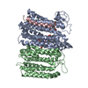


 PDBj
PDBj
 Assembly
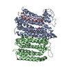

 Mass: 618.503 Da / Num. of mol.: 2 / Source method: obtained synthetically / Formula: C34H34FeN4O4
 Mass: 632.487 Da / Num. of mol.: 1 / Source method: obtained synthetically / Formula: C34H32FeN4O5
 Sample preparation
 / Beamline: X10SA / Wavelength: 1.73829 Å
 Processing