 Movie
Movie Controller

[English] 日本語
 Yorodumi
- PDB-4imh: Crystal Structure of Cytoplasmic Heme Binding Protein, PhuS, from... -
+ Open data

- Basic information
| Entry | Database: PDB / ID: 4imh | ||||||
|---|---|---|---|---|---|---|---|
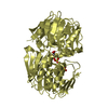
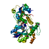
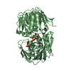
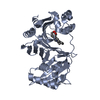
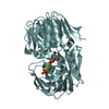
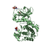
| Title | Crystal Structure of Cytoplasmic Heme Binding Protein, PhuS, from Pseudomonas aeruginosa | ||||||
 Components Components | Hemin degrading factor | ||||||
 Keywords Keywords | METAL TRANSPORT / TRANSPORT PROTEIN / Heme Transport / Heme Oxygenase | ||||||
| Function / homology |  Function and homology information Function and homology information | ||||||
| Biological species |   Pseudomonas aeruginosa (bacteria) Pseudomonas aeruginosa (bacteria) | ||||||
| Method |  X-RAY DIFFRACTION / SYNCHROTRON / MOLECULAR REPLACEMENT / Resolution: 1.976 Å X-RAY DIFFRACTION / SYNCHROTRON / MOLECULAR REPLACEMENT / Resolution: 1.976 Å | ||||||
 Authors Authors | Tripathi, S.M. / Poulos, T.L. | ||||||
 Citation Citation | Journal: J.Inorg.Biochem. / Year: 2013 Title: Crystal structure of the Pseudomonas aeruginosa cytoplasmic heme binding protein, Apo-PhuS. Authors: Tripathi, S. / O'Neill, M.J. / Wilks, A. / Poulos, T.L. | ||||||
| History |
|
- Structure visualization
| Structure viewer | Molecule: MolmilJmol/JSmol |
|---|
- Downloads & links
-Download
| PDBx/mmCIF format | 4imh.cif.gz | 403.3 KB | Display | PDBx/mmCIF format |
|---|---|---|---|---|
| PDB format | pdb4imh.ent.gz | 337.7 KB | Display | PDB format |
| PDBx/mmJSON format | 4imh.json.gz | Tree view | PDBx/mmJSON format | |
| Others |  Other downloads Other downloads |
-Validation report
| Arichive directory | https://data.pdbj.org/pub/pdb/validation_reports/im/4imhftp://data.pdbj.org/pub/pdb/validation_reports/im/4imh | HTTPS FTP |
|---|
-Related structure data
| Related structure data |  2hq2 S: Starting model for refinement |
|---|---|
| Similar structure data |


-Links
 PDBj
PDBj- Assembly
| Deposited unit | 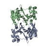
| ||||||||
|---|---|---|---|---|---|---|---|---|---|
| 1 | 
| ||||||||
| 2 | 
| ||||||||
| Unit cell |
|
-Components
| #1: Protein | Mass: 39962.191 Da / Num. of mol.: 2 Source method: isolated from a genetically manipulated source Source: (gene. exp.) Pseudomonas aeruginosa (bacteria) / Gene: phuS / Plasmid: pET28a / Production host: #2: Water | ChemComp-HOH / |  Mass: 18.015 Da / Num. of mol.: 371 / Source method: isolated from a natural source / Formula: H2O Mass: 18.015 Da / Num. of mol.: 371 / Source method: isolated from a natural source / Formula: H2O |
|---|
-Experimental details
-Experiment
| Experiment | Method: X-RAY DIFFRACTION / Number of used crystals: 1 |
|---|
- Sample preparation
| Crystal | Density Matthews: 2.34 Å3/Da / Density % sol: 47.42 % |
|---|---|
| Crystal grow | Temperature: 295 K / Method: vapor diffusion, hanging drop / pH: 7.5 Details: 0.2M MgCl2, 0.1M HEPES(7.5), 25% polyacrylic acid 5100, VAPOR DIFFUSION, HANGING DROP, temperature 295K |
-Data collection
| Diffraction | Mean temperature: 100 K |
|---|---|
| Diffraction source | Source: SYNCHROTRON / Site: SSRL  / Beamline: BL7-1 / Wavelength: 1.09 Å / Beamline: BL7-1 / Wavelength: 1.09 Å |
| Detector | Type: ADSC QUANTUM 315 / Detector: CCD / Date: Jul 5, 2011 |
| Radiation | Monochromator: GRAPHITE / Protocol: SINGLE WAVELENGTH / Monochromatic (M) / Laue (L): M / Scattering type: x-ray |
| Radiation wavelength | Wavelength: 1.09 Å / Relative weight: 1 |
| Reflection | Resolution: 1.976→46.93 Å / Num. obs: 51833 / % possible obs: 98.9 % / Observed criterion σ(I): -3 / Redundancy: 5.3 % / Rmerge(I) obs: 0.059 / Net I/σ(I): 15.7 |
| Reflection shell | Resolution: 1.976→2.08 Å / Redundancy: 5.4 % / Rmerge(I) obs: 0.24 / Mean I/σ(I) obs: 5.6 / % possible all: 100 |
- Processing
| Software |
| ||||||||||||||||||||||||||||||||||||||||||||||||||||||||||||||||||||||||||||||||||||||||||||||||||||||||||||||||||||||||||||||||||||||||||||
|---|---|---|---|---|---|---|---|---|---|---|---|---|---|---|---|---|---|---|---|---|---|---|---|---|---|---|---|---|---|---|---|---|---|---|---|---|---|---|---|---|---|---|---|---|---|---|---|---|---|---|---|---|---|---|---|---|---|---|---|---|---|---|---|---|---|---|---|---|---|---|---|---|---|---|---|---|---|---|---|---|---|---|---|---|---|---|---|---|---|---|---|---|---|---|---|---|---|---|---|---|---|---|---|---|---|---|---|---|---|---|---|---|---|---|---|---|---|---|---|---|---|---|---|---|---|---|---|---|---|---|---|---|---|---|---|---|---|---|---|---|---|
| Refinement | Method to determine structure: MOLECULAR REPLACEMENT Starting model: PDB ENTRY 2HQ2 2hq2 Resolution: 1.976→41.978 Å / SU ML: 0.21 / σ(F): 1.35 / Phase error: 23.38 / Stereochemistry target values: ML
| ||||||||||||||||||||||||||||||||||||||||||||||||||||||||||||||||||||||||||||||||||||||||||||||||||||||||||||||||||||||||||||||||||||||||||||
| Solvent computation | Shrinkage radii: 0.9 Å / VDW probe radii: 1.11 Å / Solvent model: FLAT BULK SOLVENT MODEL | ||||||||||||||||||||||||||||||||||||||||||||||||||||||||||||||||||||||||||||||||||||||||||||||||||||||||||||||||||||||||||||||||||||||||||||
| Refinement step | Cycle: LAST / Resolution: 1.976→41.978 Å
| ||||||||||||||||||||||||||||||||||||||||||||||||||||||||||||||||||||||||||||||||||||||||||||||||||||||||||||||||||||||||||||||||||||||||||||
| Refine LS restraints |
| ||||||||||||||||||||||||||||||||||||||||||||||||||||||||||||||||||||||||||||||||||||||||||||||||||||||||||||||||||||||||||||||||||||||||||||
| LS refinement shell |
| ||||||||||||||||||||||||||||||||||||||||||||||||||||||||||||||||||||||||||||||||||||||||||||||||||||||||||||||||||||||||||||||||||||||||||||
| Refinement TLS params. | Method: refined / Refine-ID: X-RAY DIFFRACTION
| ||||||||||||||||||||||||||||||||||||||||||||||||||||||||||||||||||||||||||||||||||||||||||||||||||||||||||||||||||||||||||||||||||||||||||||
| Refinement TLS group |
|
