 Movie
Movie Controller


+ Open data

- Basic information
| Entry | Database: PDB / ID: 7opb | ||||||
|---|---|---|---|---|---|---|---|
| Title | IL7R in complex with an antagonist | ||||||
 Components Components |
| ||||||
 Keywords Keywords | PROTEIN BINDING / protein binder / IL7R / antagonist | ||||||
| Function / homology |  Function and homology information Function and homology informationinterleukin-7 receptor activity / negative regulation of T cell mediated cytotoxicity / regulation of DNA recombination / positive regulation of T cell differentiation in thymus / positive regulation of receptor signaling pathway via STAT / interleukin-7-mediated signaling pathway / negative regulation of T cell apoptotic process / cellular homeostasis / cytokine receptor activity / regulation of cell size ...interleukin-7 receptor activity / negative regulation of T cell mediated cytotoxicity / regulation of DNA recombination / positive regulation of T cell differentiation in thymus / positive regulation of receptor signaling pathway via STAT / interleukin-7-mediated signaling pathway / negative regulation of T cell apoptotic process / cellular homeostasis / cytokine receptor activity / regulation of cell size / B cell homeostasis / T cell homeostasis / B cell proliferation / hemopoiesis / lymph node development / antigen binding / Interleukin-7 signaling / T cell mediated cytotoxicity / positive regulation of receptor signaling pathway via JAK-STAT / clathrin-coated endocytic vesicle membrane / cell morphogenesis / T cell differentiation in thymus / Cargo recognition for clathrin-mediated endocytosis / Clathrin-mediated endocytosis / gene expression / cell surface receptor signaling pathway / defense response to Gram-positive bacterium / immune response / external side of plasma membrane / positive regulation of cell population proliferation / positive regulation of gene expression / signal transduction / extracellular region / nucleoplasm / plasma membrane / cytosol Similarity search - Function | ||||||
| Biological species |  Homo sapiens (human) Homo sapiens (human) | ||||||
| Method |  X-RAY DIFFRACTION / SYNCHROTRON / MOLECULAR REPLACEMENT / Resolution: 2.144 Å X-RAY DIFFRACTION / SYNCHROTRON / MOLECULAR REPLACEMENT / Resolution: 2.144 Å | ||||||
 Authors Authors | Markovic, I. / Verschueren, K.H.G. / Verstraete, K. / Savvides, S.N. | ||||||
 Citation Citation | Journal: Nature / Year: 2022 Title: Design of protein-binding proteins from the target structure alone. Authors: Cao, L. / Coventry, B. / Goreshnik, I. / Huang, B. / Sheffler, W. / Park, J.S. / Jude, K.M. / Markovic, I. / Kadam, R.U. / Verschueren, K.H.G. / Verstraete, K. / Walsh, S.T.R. / Bennett, N. ...Authors: Cao, L. / Coventry, B. / Goreshnik, I. / Huang, B. / Sheffler, W. / Park, J.S. / Jude, K.M. / Markovic, I. / Kadam, R.U. / Verschueren, K.H.G. / Verstraete, K. / Walsh, S.T.R. / Bennett, N. / Phal, A. / Yang, A. / Kozodoy, L. / DeWitt, M. / Picton, L. / Miller, L. / Strauch, E.M. / DeBouver, N.D. / Pires, A. / Bera, A.K. / Halabiya, S. / Hammerson, B. / Yang, W. / Bernard, S. / Stewart, L. / Wilson, I.A. / Ruohola-Baker, H. / Schlessinger, J. / Lee, S. / Savvides, S.N. / Garcia, K.C. / Baker, D. | ||||||
| History |
|
- Structure visualization
| Structure viewer | Molecule: MolmilJmol/JSmol |
|---|
- Downloads & links
-Download
| PDBx/mmCIF format | 7opb.cif.gz | 171.5 KB | Display | PDBx/mmCIF format |
|---|---|---|---|---|
| PDB format | pdb7opb.ent.gz | 136.1 KB | Display | PDB format |
| PDBx/mmJSON format | 7opb.json.gz | Tree view | PDBx/mmJSON format | |
| Others |  Other downloads Other downloads |
-Validation report
| Arichive directory | https://data.pdbj.org/pub/pdb/validation_reports/op/7opbftp://data.pdbj.org/pub/pdb/validation_reports/op/7opb | HTTPS FTP |
|---|
-Related structure data
| Related structure data | 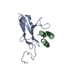 7n1jC 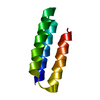 7n1kC  7n3tC 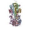 7rdhC 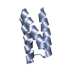 7s5bC 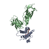 3di2S S: Starting model for refinement C: citing same article ( |
|---|---|
| Similar structure data |
-Links
 PDBj
PDBj


- Assembly
| Deposited unit | 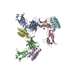
| ||||||||
|---|---|---|---|---|---|---|---|---|---|
| 1 | 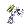
| ||||||||
| 2 | 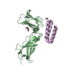
| ||||||||
| 3 | 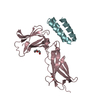
| ||||||||
| Unit cell |
|
-Components
-Protein , 2 types, 6 molecules ABCDEF
| #1: Protein | Mass: 22862.211 Da / Num. of mol.: 3 Source method: isolated from a genetically manipulated source Source: (gene. exp.) Homo sapiens (human) / Gene: IL7R / Production host: #2: Protein | Mass: 6443.422 Da / Num. of mol.: 3 Source method: isolated from a genetically manipulated source Source: (gene. exp.) |
|---|
-Non-polymers , 4 types, 315 molecules 


| #3: Chemical | ChemComp-PGE /  Mass: 150.173 Da / Num. of mol.: 1 / Source method: obtained synthetically / Formula: C6H14O4 Mass: 150.173 Da / Num. of mol.: 1 / Source method: obtained synthetically / Formula: C6H14O4 | ||||
|---|---|---|---|---|---|
| #4: Chemical |  Mass: 62.068 Da / Num. of mol.: 3 / Source method: obtained synthetically / Formula: C2H6O2 Mass: 62.068 Da / Num. of mol.: 3 / Source method: obtained synthetically / Formula: C2H6O2#5: Chemical |  Mass: 106.120 Da / Num. of mol.: 2 / Source method: obtained synthetically / Formula: C4H10O3 Mass: 106.120 Da / Num. of mol.: 2 / Source method: obtained synthetically / Formula: C4H10O3#6: Water | ChemComp-HOH / | Mass: 18.015 Da / Num. of mol.: 309 / Source method: isolated from a natural source / Formula: H2O |
-Details
| Has ligand of interest | N |
|---|---|
| Has protein modification | Y |
-Experimental details
-Experiment
| Experiment | Method: X-RAY DIFFRACTION / Number of used crystals: 1 |
|---|
- Sample preparation
| Crystal | Density Matthews: 3.38 Å3/Da / Density % sol: 63.6 % |
|---|---|
| Crystal grow | Temperature: 293.15 K / Method: vapor diffusion, sitting drop / pH: 5.5 Details: 0,1 M Phosphate/Citrate, 25% v/v PEG Smear Low Cryoprotected with 25% PEG-400 |
-Data collection
| Diffraction | Mean temperature: 100 K / Serial crystal experiment: N |
|---|---|
| Diffraction source | Source: SYNCHROTRON / Site: ESRF  / Beamline: ID23-1 / Wavelength: 0.87313 Å / Beamline: ID23-1 / Wavelength: 0.87313 Å |
| Detector | Type: DECTRIS PILATUS3 X 2M / Detector: PIXEL / Date: Feb 21, 2021 |
| Radiation | Protocol: SINGLE WAVELENGTH / Monochromatic (M) / Laue (L): M / Scattering type: x-ray |
| Radiation wavelength | Wavelength: 0.87313 Å / Relative weight: 1 |
| Reflection | Resolution: 2.144→43.96 Å / Num. obs: 62839 / % possible obs: 99.6 % / Redundancy: 8.19 % / Biso Wilson estimate: 34.17 Å2 / CC1/2: 0.993 / Rpim(I) all: 0.08 / Rrim(I) all: 0.204 / Χ2: 1.02 / Net I/av σ(I): 6.92 / Net I/σ(I): 6.92 |
| Reflection shell | Resolution: 2.144→2.27 Å / Mean I/σ(I) obs: 1.04 / Num. unique obs: 9978 / CC1/2: 0.442 / Rpim(I) all: 0.868 / Rrim(I) all: 1.94 / Χ2: 0.98 / % possible all: 97.6 |
- Processing
| Software |
| ||||||||||||||||||||||||||||||||||||||||||||||||||||||||||||
|---|---|---|---|---|---|---|---|---|---|---|---|---|---|---|---|---|---|---|---|---|---|---|---|---|---|---|---|---|---|---|---|---|---|---|---|---|---|---|---|---|---|---|---|---|---|---|---|---|---|---|---|---|---|---|---|---|---|---|---|---|---|
| Refinement | Method to determine structure: MOLECULAR REPLACEMENT Starting model: 3DI2 Resolution: 2.144→43.96 Å / Cor.coef. Fo:Fc: 0.939 / Cor.coef. Fo:Fc free: 0.933 / SU R Cruickshank DPI: 0.175 / Cross valid method: THROUGHOUT / SU R Blow DPI: 0.171 / SU Rfree Blow DPI: 0.142 / SU Rfree Cruickshank DPI: 0.146
| ||||||||||||||||||||||||||||||||||||||||||||||||||||||||||||
| Displacement parameters | Biso mean: 58.66 Å2
| ||||||||||||||||||||||||||||||||||||||||||||||||||||||||||||
| Refine analyze | Luzzati coordinate error obs: 0.33 Å | ||||||||||||||||||||||||||||||||||||||||||||||||||||||||||||
| Refinement step | Cycle: LAST / Resolution: 2.144→43.96 Å
| ||||||||||||||||||||||||||||||||||||||||||||||||||||||||||||
| Refine LS restraints |
| ||||||||||||||||||||||||||||||||||||||||||||||||||||||||||||
| LS refinement shell | Resolution: 2.144→2.16 Å
|
