 Movie
Movie Controller

[English] 日本語
 Yorodumi
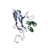
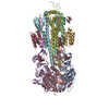
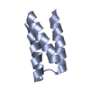
- PDB-7n1k: Crystal structure of a de novo-designed mini-protein targeting FGFR -
+ Open data

- Basic information
| Entry | Database: PDB / ID: 7n1k | ||||||
|---|---|---|---|---|---|---|---|
| Title | Crystal structure of a de novo-designed mini-protein targeting FGFR | ||||||
 Components Components | Binder | ||||||
 Keywords Keywords | DE NOVO PROTEIN / Binder | ||||||
| Biological species | synthetic construct (others) | ||||||
| Method |  X-RAY DIFFRACTION / SYNCHROTRON / MOLECULAR REPLACEMENT / Resolution: 3.01 Å X-RAY DIFFRACTION / SYNCHROTRON / MOLECULAR REPLACEMENT / Resolution: 3.01 Å | ||||||
 Authors Authors | Park, J.S. / Lee, S. | ||||||
| Funding support | 1items
| ||||||
 Citation Citation | Journal: Nature / Year: 2022 Title: Design of protein-binding proteins from the target structure alone. Authors: Cao, L. / Coventry, B. / Goreshnik, I. / Huang, B. / Sheffler, W. / Park, J.S. / Jude, K.M. / Markovic, I. / Kadam, R.U. / Verschueren, K.H.G. / Verstraete, K. / Walsh, S.T.R. / Bennett, N. ...Authors: Cao, L. / Coventry, B. / Goreshnik, I. / Huang, B. / Sheffler, W. / Park, J.S. / Jude, K.M. / Markovic, I. / Kadam, R.U. / Verschueren, K.H.G. / Verstraete, K. / Walsh, S.T.R. / Bennett, N. / Phal, A. / Yang, A. / Kozodoy, L. / DeWitt, M. / Picton, L. / Miller, L. / Strauch, E.M. / DeBouver, N.D. / Pires, A. / Bera, A.K. / Halabiya, S. / Hammerson, B. / Yang, W. / Bernard, S. / Stewart, L. / Wilson, I.A. / Ruohola-Baker, H. / Schlessinger, J. / Lee, S. / Savvides, S.N. / Garcia, K.C. / Baker, D. | ||||||
| History |
|
- Structure visualization
| Structure viewer | Molecule:  MolmilJmol/JSmol MolmilJmol/JSmol |
|---|
- Downloads & links
-Download
| PDBx/mmCIF format | 7n1k.cif.gz | 26 KB | Display | PDBx/mmCIF format |
|---|---|---|---|---|
| PDB format | pdb7n1k.ent.gz | 12 KB | Display | PDB format |
| PDBx/mmJSON format | 7n1k.json.gz | Tree view | PDBx/mmJSON format | |
| Others |  Other downloads Other downloads |
-Validation report
| Arichive directory | https://data.pdbj.org/pub/pdb/validation_reports/n1/7n1kftp://data.pdbj.org/pub/pdb/validation_reports/n1/7n1k | HTTPS FTP |
|---|
-Related structure data


-Links
 PDBj
PDBj
- Assembly
| Deposited unit | 
| ||||||||||||
|---|---|---|---|---|---|---|---|---|---|---|---|---|---|
| 1 |
| ||||||||||||
| Unit cell |
|
-Components
| #1: Protein | Mass: 7535.809 Da / Num. of mol.: 1 Source method: isolated from a genetically manipulated source Source: (gene. exp.) synthetic construct (others) / Production host:  |
|---|---|
| Has ligand of interest | N |
-Experimental details
-Experiment
| Experiment | Method: X-RAY DIFFRACTION / Number of used crystals: 1 |
|---|
- Sample preparation
| Crystal | Density Matthews: 2.49 Å3/Da / Density % sol: 50.58 % |
|---|---|
| Crystal grow | Temperature: 295.15 K / Method: vapor diffusion, hanging drop Details: 1,6-hexanediol, 1-butanol, 1,2-propanediol, 2-propanol, 1,4-butanediol, 1,3-Propanediol, Tris-BICINE (pH 8.5), MPD, PEG 1000, PEG 3350 |
-Data collection
| Diffraction | Mean temperature: 100 K / Serial crystal experiment: N |
|---|---|
| Diffraction source | Source: SYNCHROTRON / Site: APS  / Beamline: 24-ID-E / Wavelength: 0.97918 Å / Beamline: 24-ID-E / Wavelength: 0.97918 Å |
| Detector | Type: DECTRIS EIGER X 16M / Detector: PIXEL / Date: Feb 12, 2021 |
| Radiation | Protocol: SINGLE WAVELENGTH / Monochromatic (M) / Laue (L): M / Scattering type: x-ray |
| Radiation wavelength | Wavelength: 0.97918 Å / Relative weight: 1 |
| Reflection | Resolution: 3.01→50 Å / Num. obs: 1644 / % possible obs: 93.8 % / Redundancy: 5.5 % / CC1/2: 0.999 / Net I/σ(I): 17.85 |
| Reflection shell | Resolution: 3.01→3.19 Å / Num. unique obs: 247 / CC1/2: 0.99 |
- Processing
| Software |
| ||||||||||||||||||||||||
|---|---|---|---|---|---|---|---|---|---|---|---|---|---|---|---|---|---|---|---|---|---|---|---|---|---|
| Refinement | Method to determine structure: MOLECULAR REPLACEMENT Starting model: PREDICTED MODEL Resolution: 3.01→42.48 Å / SU ML: 0 / Cross valid method: FREE R-VALUE / σ(F): 1.42 / Phase error: 24.1369 Stereochemistry target values: GeoStd + Monomer Library + CDL v1.2
| ||||||||||||||||||||||||
| Solvent computation | Shrinkage radii: 0.9 Å / VDW probe radii: 1.11 Å / Solvent model: FLAT BULK SOLVENT MODEL | ||||||||||||||||||||||||
| Displacement parameters | Biso mean: 64.05 Å2 | ||||||||||||||||||||||||
| Refinement step | Cycle: LAST / Resolution: 3.01→42.48 Å
| ||||||||||||||||||||||||
| Refine LS restraints |
| ||||||||||||||||||||||||
| LS refinement shell | Resolution: 3.01→42.48 Å
|
