| 登録情報 | データベース: PDB / ID: 7n1j
|
|---|
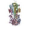
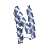
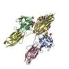
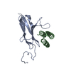
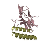
| タイトル | Crystal structure of FGFR4 domain 3 in complex with a de novo-designed mini-binder |
|---|
 要素 要素 | - Binder
- Fibroblast growth factor receptor 4
|
|---|
 キーワード キーワード | SIGNALING PROTEIN / Receptor tyrosine kinase / Complex / Binder / MEMBRANE PROTEIN |
|---|
| 機能・相同性 |  機能・相同性情報 機能・相同性情報
FGFR4 mutant receptor activation / betaKlotho-mediated ligand binding / positive regulation of catalytic activity / regulation of extracellular matrix disassembly / phosphate ion homeostasis / regulation of bile acid biosynthetic process / FGFR4 ligand binding and activation / Phospholipase C-mediated cascade; FGFR4 / fibroblast growth factor receptor activity / positive regulation of DNA biosynthetic process ...FGFR4 mutant receptor activation / betaKlotho-mediated ligand binding / positive regulation of catalytic activity / regulation of extracellular matrix disassembly / phosphate ion homeostasis / regulation of bile acid biosynthetic process / FGFR4 ligand binding and activation / Phospholipase C-mediated cascade; FGFR4 / fibroblast growth factor receptor activity / positive regulation of DNA biosynthetic process / PI-3K cascade:FGFR4 / positive regulation of proteolysis / regulation of lipid metabolic process / fibroblast growth factor binding / fibroblast growth factor receptor signaling pathway / PI3K Cascade / transport vesicle / SHC-mediated cascade:FGFR4 / Signaling by FGFR4 in disease / FRS-mediated FGFR4 signaling / peptidyl-tyrosine phosphorylation / cholesterol homeostasis / Negative regulation of FGFR4 signaling / receptor protein-tyrosine kinase / Constitutive Signaling by Aberrant PI3K in Cancer / protein autophosphorylation / PIP3 activates AKT signaling / cell migration / heparin binding / glucose homeostasis / PI5P, PP2A and IER3 Regulate PI3K/AKT Signaling / RAF/MAP kinase cascade / positive regulation of ERK1 and ERK2 cascade / signaling receptor complex / endosome / positive regulation of cell population proliferation / positive regulation of gene expression / Golgi apparatus / endoplasmic reticulum / extracellular region / ATP binding / plasma membrane類似検索 - 分子機能 Fibroblast growth factor receptor family / Immunoglobulin domain / Immunoglobulin I-set / Immunoglobulin I-set domain / : / Immunoglobulin subtype 2 / Immunoglobulin C-2 Type / Tyrosine-protein kinase, catalytic domain / Tyrosine kinase, catalytic domain / Tyrosine protein kinases specific active-site signature. ...Fibroblast growth factor receptor family / Immunoglobulin domain / Immunoglobulin I-set / Immunoglobulin I-set domain / : / Immunoglobulin subtype 2 / Immunoglobulin C-2 Type / Tyrosine-protein kinase, catalytic domain / Tyrosine kinase, catalytic domain / Tyrosine protein kinases specific active-site signature. / Immunoglobulin subtype / Immunoglobulin / Tyrosine-protein kinase, active site / Protein tyrosine and serine/threonine kinase / Serine-threonine/tyrosine-protein kinase, catalytic domain / Ig-like domain profile. / Immunoglobulin-like domain / Immunoglobulin-like domain superfamily / Protein kinase, ATP binding site / Protein kinases ATP-binding region signature. / Immunoglobulin-like fold / Immunoglobulins / Protein kinase domain profile. / Protein kinase domain / Protein kinase-like domain superfamily / Immunoglobulin-like / Sandwich / Mainly Beta類似検索 - ドメイン・相同性 |
|---|
| 生物種 |  Homo sapiens (ヒト) Homo sapiens (ヒト)
synthetic construct (人工物) |
|---|
| 手法 |  X線回折 / シンクロトロン / 分子置換 / 解像度: 2.99 Å X線回折 / シンクロトロン / 分子置換 / 解像度: 2.99 Å |
|---|
 データ登録者 データ登録者 | Park, J.S. / Lee, S. |
|---|
| 資金援助 | 1件 |
|---|
 引用 引用 | ジャーナル: Nature / 年: 2022
タイトル: Design of protein-binding proteins from the target structure alone.
著者: Cao, L. / Coventry, B. / Goreshnik, I. / Huang, B. / Sheffler, W. / Park, J.S. / Jude, K.M. / Markovic, I. / Kadam, R.U. / Verschueren, K.H.G. / Verstraete, K. / Walsh, S.T.R. / Bennett, N. / ...著者: Cao, L. / Coventry, B. / Goreshnik, I. / Huang, B. / Sheffler, W. / Park, J.S. / Jude, K.M. / Markovic, I. / Kadam, R.U. / Verschueren, K.H.G. / Verstraete, K. / Walsh, S.T.R. / Bennett, N. / Phal, A. / Yang, A. / Kozodoy, L. / DeWitt, M. / Picton, L. / Miller, L. / Strauch, E.M. / DeBouver, N.D. / Pires, A. / Bera, A.K. / Halabiya, S. / Hammerson, B. / Yang, W. / Bernard, S. / Stewart, L. / Wilson, I.A. / Ruohola-Baker, H. / Schlessinger, J. / Lee, S. / Savvides, S.N. / Garcia, K.C. / Baker, D. |
|---|
| 履歴 | | 登録 | 2021年5月27日 | 登録サイト: RCSB / 処理サイト: RCSB |
|---|
| 改定 1.0 | 2022年4月6日 | Provider: repository / タイプ: Initial release |
|---|
| 改定 1.1 | 2022年5月18日 | Group: Database references / カテゴリ: citation / citation_author / Item: _citation.title |
|---|
| 改定 1.2 | 2022年5月25日 | Group: Database references / カテゴリ: citation
Item: _citation.journal_volume / _citation.page_first / _citation.page_last |
|---|
| 改定 1.3 | 2023年10月18日 | Group: Data collection / Refinement description
カテゴリ: chem_comp_atom / chem_comp_bond / pdbx_initial_refinement_model |
|---|
| 改定 1.4 | 2024年10月30日 | Group: Structure summary
カテゴリ: pdbx_entry_details / pdbx_modification_feature |
|---|
|
|---|
 ムービー
ムービー コントローラー


 データを開く
 基本情報
基本情報 構造の表示
構造の表示 ダウンロードとリンク
 その他のダウンロード
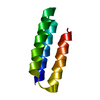


 PDBj
PDBj


 集合体
集合体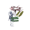

 試料調製
試料調製 / ビームライン: 24-ID-E / 波長: 0.97918 Å
 解析
解析