 Movie
Movie Controller

[English] 日本語
 Yorodumi
- PDB-7op7: Bacteroides thetaiotaomicron mannosidase GH2 with beta-manno-conf... -
+ Open data

- Basic information
| Entry | Database: PDB / ID: 7op7 | ||||||||||||
|---|---|---|---|---|---|---|---|---|---|---|---|---|---|
| Title | Bacteroides thetaiotaomicron mannosidase GH2 with beta-manno-configured N-alkyl cyclophellitol aziridine | ||||||||||||
 Components Components | Beta-mannosidase | ||||||||||||
 Keywords Keywords | HYDROLASE / mannosidase / cyclophellitol aziridine | ||||||||||||
| Function / homology |  Function and homology information Function and homology informationbeta-mannosidase / beta-mannosidase activity / glycoprotein catabolic process / carbohydrate metabolic process / extracellular region Similarity search - Function | ||||||||||||
| Biological species |  Bacteroides thetaiotaomicron VPI-5482 (bacteria) Bacteroides thetaiotaomicron VPI-5482 (bacteria) | ||||||||||||
| Method |  X-RAY DIFFRACTION / SYNCHROTRON / MOLECULAR REPLACEMENT / Resolution: 1.85 Å X-RAY DIFFRACTION / SYNCHROTRON / MOLECULAR REPLACEMENT / Resolution: 1.85 Å | ||||||||||||
 Authors Authors | McGregor, N.G.S. / Beenakker, T.J.M. / Kuo, C. / Wong, C. / Offen, W.A. / Armstrong, Z. / Codee, J.D.C. / Aerts, J.M.F.G. / Florea, B.I. / Overkleeft, H.S. / Davies, G.J. | ||||||||||||
| Funding support |  United Kingdom, European Union, 3items United Kingdom, European Union, 3items
| ||||||||||||
 Citation Citation | Journal: Org.Biomol.Chem. / Year: 2022 Title: Synthesis of broad-specificity activity-based probes for exo -beta-mannosidases. Authors: McGregor, N.G.S. / Kuo, C.L. / Beenakker, T.J.M. / Wong, C.S. / Offen, W.A. / Armstrong, Z. / Florea, B.I. / Codee, J.D.C. / Overkleeft, H.S. / Aerts, J.M.F.G. / Davies, G.J. | ||||||||||||
| History |
|
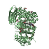
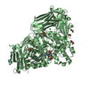
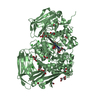
- Structure visualization
| Structure viewer | Molecule: MolmilJmol/JSmol |
|---|
- Downloads & links
-Download
| PDBx/mmCIF format | 7op7.cif.gz | 382.6 KB | Display | PDBx/mmCIF format |
|---|---|---|---|---|
| PDB format | pdb7op7.ent.gz | 301.9 KB | Display | PDB format |
| PDBx/mmJSON format | 7op7.json.gz | Tree view | PDBx/mmJSON format | |
| Others |  Other downloads Other downloads |
-Validation report
| Summary document | 7op7_validation.pdf.gz | 1007.6 KB | Display | wwPDB validaton report |
|---|---|---|---|---|
| Full document | 7op7_full_validation.pdf.gz | 1023.5 KB | Display | |
| Data in XML | 7op7_validation.xml.gz | 68.7 KB | Display | |
| Data in CIF | 7op7_validation.cif.gz | 101.6 KB | Display | |
| Arichive directory | https://data.pdbj.org/pub/pdb/validation_reports/op/7op7ftp://data.pdbj.org/pub/pdb/validation_reports/op/7op7 | HTTPS FTP |
-Related structure data
| Related structure data |  7odjC 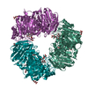 7omiC 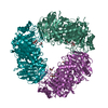 7omsC 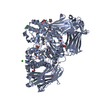 7op6C 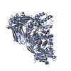 2je8S S: Starting model for refinement C: citing same article ( |
|---|---|
| Similar structure data |
-Links
 PDBj
PDBj


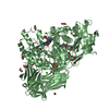
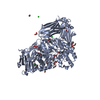
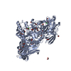
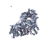
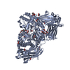
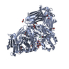
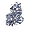
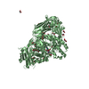
- Assembly
| Deposited unit | 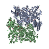
| ||||||||
|---|---|---|---|---|---|---|---|---|---|
| 1 | 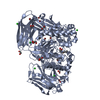
| ||||||||
| 2 | 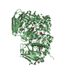
| ||||||||
| Unit cell |
|
-Components
-Protein , 1 types, 2 molecules AB
| #1: Protein | Mass: 98291.125 Da / Num. of mol.: 2 Source method: isolated from a genetically manipulated source Details: beta-mannosidase without signal peptide sequence and with C-terminal his6 tag Source: (gene. exp.) Bacteroides thetaiotaomicron VPI-5482 (bacteria)Gene: BT_0458 / Production host: |
|---|
-Non-polymers , 5 types, 889 molecules 
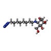


| #2: Chemical | ChemComp-EDO /  Mass: 62.068 Da / Num. of mol.: 28 / Source method: obtained synthetically / Formula: C2H6O2 Mass: 62.068 Da / Num. of mol.: 28 / Source method: obtained synthetically / Formula: C2H6O2#3: Chemical |  Mass: 330.423 Da / Num. of mol.: 2 / Source method: obtained synthetically / Formula: C15H30N4O4 / Feature type: SUBJECT OF INVESTIGATION Mass: 330.423 Da / Num. of mol.: 2 / Source method: obtained synthetically / Formula: C15H30N4O4 / Feature type: SUBJECT OF INVESTIGATION#4: Chemical | ChemComp-BR /  Mass: 79.904 Da / Num. of mol.: 13 / Source method: obtained synthetically / Formula: Br Mass: 79.904 Da / Num. of mol.: 13 / Source method: obtained synthetically / Formula: Br#5: Chemical | ChemComp-CL /  Mass: 35.453 Da / Num. of mol.: 18 / Source method: obtained synthetically / Formula: Cl Mass: 35.453 Da / Num. of mol.: 18 / Source method: obtained synthetically / Formula: Cl#6: Water | ChemComp-HOH / | Mass: 18.015 Da / Num. of mol.: 828 / Source method: isolated from a natural source / Formula: H2O |
|---|
-Details
| Has ligand of interest | Y |
|---|---|
| Has protein modification | Y |
-Experimental details
-Experiment
| Experiment | Method: X-RAY DIFFRACTION / Number of used crystals: 1 |
|---|
- Sample preparation
| Crystal | Density Matthews: 2.41 Å3/Da / Density % sol: 49 % |
|---|---|
| Crystal grow | Temperature: 291 K / Method: vapor diffusion, sitting drop / pH: 5.5 / Details: PEG 3350, sodium bromide, MES buffer |
-Data collection
| Diffraction | Mean temperature: 100 K / Serial crystal experiment: N |
|---|---|
| Diffraction source | Source: SYNCHROTRON / Site: Diamond / Beamline: I03 / Wavelength: 0.97629 Å |
| Detector | Type: DECTRIS EIGER2 X 16M / Detector: PIXEL / Date: Apr 25, 2021 |
| Radiation | Protocol: SINGLE WAVELENGTH / Monochromatic (M) / Laue (L): M / Scattering type: x-ray |
| Radiation wavelength | Wavelength: 0.97629 Å / Relative weight: 1 |
| Reflection | Resolution: 1.85→67.66 Å / Num. obs: 158303 / % possible obs: 99.9 % / Redundancy: 5.5 % / CC1/2: 0.997 / Rpim(I) all: 0.053 / Net I/σ(I): 8.3 |
| Reflection shell | Resolution: 1.85→1.88 Å / Mean I/σ(I) obs: 0.7 / Num. unique obs: 7816 / CC1/2: 0.5 / Rpim(I) all: 1.004 / % possible all: 99.8 |
- Processing
| Software |
| ||||||||||||||||||||||||||||||||||||||||||||||||||||||||||||
|---|---|---|---|---|---|---|---|---|---|---|---|---|---|---|---|---|---|---|---|---|---|---|---|---|---|---|---|---|---|---|---|---|---|---|---|---|---|---|---|---|---|---|---|---|---|---|---|---|---|---|---|---|---|---|---|---|---|---|---|---|---|
| Refinement | Method to determine structure: MOLECULAR REPLACEMENT Starting model: 2JE8.pdb Resolution: 1.85→67.66 Å / Cor.coef. Fo:Fc: 0.964 / Cor.coef. Fo:Fc free: 0.941 / SU B: 5.432 / SU ML: 0.147 / Cross valid method: THROUGHOUT / σ(F): 0 / ESU R: 0.145 / ESU R Free: 0.145 / Stereochemistry target values: MAXIMUM LIKELIHOOD Details: HYDROGENS HAVE BEEN ADDED IN THE RIDING POSITIONS U VALUES : REFINED INDIVIDUALLY THE ALKYL CHAIN OF THE LIGAND IN MOLECULE A IS MODELLED PARTLY, WITH SOME ATOMS AT PARTIAL OCCUPANCY. THE ...Details: HYDROGENS HAVE BEEN ADDED IN THE RIDING POSITIONS U VALUES : REFINED INDIVIDUALLY THE ALKYL CHAIN OF THE LIGAND IN MOLECULE A IS MODELLED PARTLY, WITH SOME ATOMS AT PARTIAL OCCUPANCY. THE LIGAND IN MOLECULE B IS MODELLED AT HALF OCCUPANCY, AND AN ETHYLENE GLYCOL AT HALF OCCUPANCY IS ALSO BOUND IN THE ACTIVE SITE.
| ||||||||||||||||||||||||||||||||||||||||||||||||||||||||||||
| Solvent computation | Ion probe radii: 0.8 Å / Shrinkage radii: 0.8 Å / VDW probe radii: 1.2 Å / Solvent model: MASK | ||||||||||||||||||||||||||||||||||||||||||||||||||||||||||||
| Displacement parameters | Biso max: 94.07 Å2 / Biso mean: 32.299 Å2 / Biso min: 12.64 Å2
| ||||||||||||||||||||||||||||||||||||||||||||||||||||||||||||
| Refinement step | Cycle: final / Resolution: 1.85→67.66 Å
| ||||||||||||||||||||||||||||||||||||||||||||||||||||||||||||
| Refine LS restraints |
| ||||||||||||||||||||||||||||||||||||||||||||||||||||||||||||
| LS refinement shell | Resolution: 1.85→1.898 Å / Rfactor Rfree error: 0 / Total num. of bins used: 20
|
