 Movie
Movie Controller


+ Open data

- Basic information
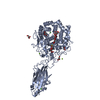
| Entry | Database: PDB / ID: 7onw | ||||||
|---|---|---|---|---|---|---|---|
| Title | Crystal structure of PBP3 from E. coli in complex with AIC499 | ||||||
 Components Components | Peptidoglycan D,D-transpeptidase FtsI | ||||||
 Keywords Keywords | PROTEIN BINDING / monobactam / PBP3 / peptidoglycan synthesis / drug complex | ||||||
| Function / homology |  Function and homology information Function and homology informationdivisome complex / peptidoglycan glycosyltransferase activity / serine-type D-Ala-D-Ala carboxypeptidase / serine-type D-Ala-D-Ala carboxypeptidase activity / division septum assembly / FtsZ-dependent cytokinesis / cell division site / penicillin binding / peptidoglycan biosynthetic process / cell wall organization ...divisome complex / peptidoglycan glycosyltransferase activity / serine-type D-Ala-D-Ala carboxypeptidase / serine-type D-Ala-D-Ala carboxypeptidase activity / division septum assembly / FtsZ-dependent cytokinesis / cell division site / penicillin binding / peptidoglycan biosynthetic process / cell wall organization / regulation of cell shape / response to xenobiotic stimulus / cell division / proteolysis / plasma membrane Similarity search - Function | ||||||
| Biological species |  | ||||||
| Method |  X-RAY DIFFRACTION / SYNCHROTRON / MOLECULAR REPLACEMENT / molecular replacement / Resolution: 2.7 Å X-RAY DIFFRACTION / SYNCHROTRON / MOLECULAR REPLACEMENT / molecular replacement / Resolution: 2.7 Å | ||||||
 Authors Authors | Freischem, S. / Grimm, I. / Weiergraeber, O.H. | ||||||
 Citation Citation | Journal: Biomolecules / Year: 2021 Title: Interaction Mode of the Novel Monobactam AIC499 Targeting Penicillin Binding Protein 3 of Gram-Negative Bacteria. Authors: Freischem, S. / Grimm, I. / Lopez-Perez, A. / Willbold, D. / Klenke, B. / Vuong, C. / Dingley, A.J. / Weiergraber, O.H. | ||||||
| History |
|
- Structure visualization
| Structure viewer | Molecule: MolmilJmol/JSmol |
|---|
- Downloads & links
-Download
| PDBx/mmCIF format | 7onw.cif.gz | 222.4 KB | Display | PDBx/mmCIF format |
|---|---|---|---|---|
| PDB format | pdb7onw.ent.gz | 155.8 KB | Display | PDB format |
| PDBx/mmJSON format | 7onw.json.gz | Tree view | PDBx/mmJSON format | |
| Others |  Other downloads Other downloads |
-Validation report
| Arichive directory | https://data.pdbj.org/pub/pdb/validation_reports/on/7onwftp://data.pdbj.org/pub/pdb/validation_reports/on/7onw | HTTPS FTP |
|---|
-Related structure data
| Related structure data | 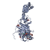 7onkC 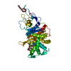 7onnC 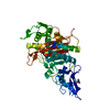 7onoC 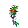 7onxC 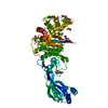 7onyC 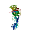 7onzC 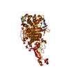 4bjpS C: citing same article ( S: Starting model for refinement |
|---|---|
| Similar structure data |


-Links
 PDBj
PDBj
- Assembly
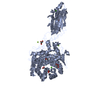
| Deposited unit | 
| ||||||||||||
|---|---|---|---|---|---|---|---|---|---|---|---|---|---|
| 1 |
| ||||||||||||
| Unit cell |
|
-Components
| #1: Protein | Mass: 61102.574 Da / Num. of mol.: 1 Source method: isolated from a genetically manipulated source Source: (gene. exp.) Strain: K12 / Gene: ftsI, pbpB, b0084, JW0082 / Production host: References: UniProt: P0AD68, serine-type D-Ala-D-Ala carboxypeptidase | ||||||||
|---|---|---|---|---|---|---|---|---|---|
| #2: Chemical | ChemComp-VL5 / (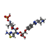  Mass: 670.715 Da / Num. of mol.: 1 / Source method: obtained synthetically / Formula: C25H34N8O10S2 / Feature type: SUBJECT OF INVESTIGATION Mass: 670.715 Da / Num. of mol.: 1 / Source method: obtained synthetically / Formula: C25H34N8O10S2 / Feature type: SUBJECT OF INVESTIGATION | ||||||||
| #3: Chemical |   Mass: 94.971 Da / Num. of mol.: 2 / Source method: obtained synthetically / Formula: PO4 Mass: 94.971 Da / Num. of mol.: 2 / Source method: obtained synthetically / Formula: PO4#4: Chemical |   Mass: 118.174 Da / Num. of mol.: 2 / Source method: obtained synthetically / Formula: C6H14O2 / Comment: precipitant*YM Mass: 118.174 Da / Num. of mol.: 2 / Source method: obtained synthetically / Formula: C6H14O2 / Comment: precipitant*YM#5: Water | ChemComp-HOH / |  Mass: 18.015 Da / Num. of mol.: 6 / Source method: isolated from a natural source / Formula: H2O Mass: 18.015 Da / Num. of mol.: 6 / Source method: isolated from a natural source / Formula: H2OHas ligand of interest | Y | Has protein modification | Y | |
-Experimental details
-Experiment
| Experiment | Method: X-RAY DIFFRACTION / Number of used crystals: 1 |
|---|
- Sample preparation
| Crystal | Density Matthews: 3.86 Å3/Da / Density % sol: 68.12 % |
|---|---|
| Crystal grow | Temperature: 293 K / Method: vapor diffusion, sitting drop / pH: 6.5 Details: 10 mg/ml PBP3, 3% dextran sulfate (M-5000), 0.1 M sodium cacodylate, 5% PEG 8000, 30% (v/v) MPD. 0.5 mM AIC499 |
-Data collection
| Diffraction | Mean temperature: 100 K / Serial crystal experiment: N |
|---|---|
| Diffraction source | Source: SYNCHROTRON / Site: ESRF  / Beamline: ID23-1 / Wavelength: 0.97242 Å / Beamline: ID23-1 / Wavelength: 0.97242 Å |
| Detector | Type: DECTRIS PILATUS 6M / Detector: PIXEL / Date: Jun 7, 2018 |
| Radiation | Protocol: SINGLE WAVELENGTH / Monochromatic (M) / Laue (L): M / Scattering type: x-ray |
| Radiation wavelength | Wavelength: 0.97242 Å / Relative weight: 1 |
| Reflection | Resolution: 2.7→48.64 Å / Num. obs: 14000 / % possible obs: 92.2 % / Redundancy: 9.4 % / CC1/2: 0.999 / Rrim(I) all: 0.101 / Net I/σ(I): 15.4 |
| Reflection shell | Resolution: 2.704→3.034 Å / Redundancy: 8.8 % / Mean I/σ(I) obs: 2 / Num. unique obs: 700 / CC1/2: 0.793 / Rrim(I) all: 1.211 / % possible all: 76.1 |
-Phasing
| Phasing | Method: molecular replacement |
|---|
- Processing
| Software |
| |||||||||||||||||||||||||||||||||||||||||||||||||||||||||||||||||||||||||||
|---|---|---|---|---|---|---|---|---|---|---|---|---|---|---|---|---|---|---|---|---|---|---|---|---|---|---|---|---|---|---|---|---|---|---|---|---|---|---|---|---|---|---|---|---|---|---|---|---|---|---|---|---|---|---|---|---|---|---|---|---|---|---|---|---|---|---|---|---|---|---|---|---|---|---|---|---|
| Refinement | Method to determine structure: MOLECULAR REPLACEMENT Starting model: 4BJP Resolution: 2.7→48.64 Å / SU ML: 0.3103 / Cross valid method: FREE R-VALUE / σ(F): 1.35 / Phase error: 31.3319 Stereochemistry target values: GeoStd + Monomer Library + CDL v1.2
| |||||||||||||||||||||||||||||||||||||||||||||||||||||||||||||||||||||||||||
| Solvent computation | Shrinkage radii: 0.9 Å / VDW probe radii: 1.11 Å / Solvent model: FLAT BULK SOLVENT MODEL | |||||||||||||||||||||||||||||||||||||||||||||||||||||||||||||||||||||||||||
| Displacement parameters | Biso mean: 82.96 Å2 | |||||||||||||||||||||||||||||||||||||||||||||||||||||||||||||||||||||||||||
| Refinement step | Cycle: LAST / Resolution: 2.7→48.64 Å
| |||||||||||||||||||||||||||||||||||||||||||||||||||||||||||||||||||||||||||
| Refine LS restraints |
| |||||||||||||||||||||||||||||||||||||||||||||||||||||||||||||||||||||||||||
| LS refinement shell |
| |||||||||||||||||||||||||||||||||||||||||||||||||||||||||||||||||||||||||||
| Refinement TLS params. | Method: refined / Refine-ID: X-RAY DIFFRACTION
| |||||||||||||||||||||||||||||||||||||||||||||||||||||||||||||||||||||||||||
| Refinement TLS group | Refine-ID: X-RAY DIFFRACTION / Auth asym-ID: A / Label asym-ID: A
|
