 Movie
Movie Controller


+ Open data

- Basic information
| Entry | Database: PDB / ID: 4bjp | ||||||
|---|---|---|---|---|---|---|---|
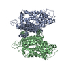
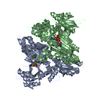
| Title | Crystal structure of E. coli penicillin binding protein 3 | ||||||
 Components Components | PENICILLIN BINDING PROTEIN TRANSPEPTIDASE DOMAIN PROTEIN | ||||||
 Keywords Keywords | TRANSFERASE | ||||||
| Function / homology | Beta-Lactamase - #330 / Beta-Lactamase / Beta-lactamase / DD-peptidase/beta-lactamase superfamily / 2-Layer Sandwich / 3-Layer(aba) Sandwich / Alpha Beta / :  Function and homology information Function and homology information | ||||||
| Biological species |  | ||||||
| Method |  X-RAY DIFFRACTION / SYNCHROTRON / MOLECULAR REPLACEMENT / Resolution: 2.5 Å X-RAY DIFFRACTION / SYNCHROTRON / MOLECULAR REPLACEMENT / Resolution: 2.5 Å | ||||||
 Authors Authors | Sauvage, E. / Joris, M. / Herman, R. / Kerff, F. / Rocaboy, M. / Charlier, P. | ||||||
 Citation Citation | Journal: Plos One / Year: 2014 Title: Crystal Structure of Penicillin-Binding Protein 3 (Pbp3) from Escherichia Coli. Authors: Sauvage, E. / Derouaux, A. / Fraipont, C. / Joris, M. / Herman, R. / Rocaboy, M. / Schloesser, M. / Dumas, J. / Kerff, F. / Nguyen-Disteche, M. / Charlier, P. | ||||||
| History |
|
- Structure visualization
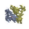
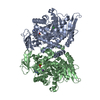
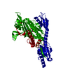
| Structure viewer | Molecule: MolmilJmol/JSmol |
|---|
- Downloads & links
-Download
| PDBx/mmCIF format | 4bjp.cif.gz | 180.4 KB | Display | PDBx/mmCIF format |
|---|---|---|---|---|
| PDB format | pdb4bjp.ent.gz | 142.2 KB | Display | PDB format |
| PDBx/mmJSON format | 4bjp.json.gz | Tree view | PDBx/mmJSON format | |
| Others |  Other downloads Other downloads |
-Validation report
| Arichive directory | https://data.pdbj.org/pub/pdb/validation_reports/bj/4bjpftp://data.pdbj.org/pub/pdb/validation_reports/bj/4bjp | HTTPS FTP |
|---|
-Related structure data
| Related structure data |  4bjqC 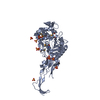 3equS C: citing same article ( S: Starting model for refinement |
|---|---|
| Similar structure data |


-Links
 PDBj
PDBj- Assembly
| Deposited unit | 
| ||||||||
|---|---|---|---|---|---|---|---|---|---|
| 1 | 
| ||||||||
| Unit cell |
|
-Components
-Protein , 1 types, 1 molecules A
| #1: Protein | Mass: 55378.129 Da / Num. of mol.: 1 / Fragment: RESIDUES 67-577 Source method: isolated from a genetically manipulated source Source: (gene. exp.) References: UniProt: J2XFH0, peptidoglycan glycosyltransferase |
|---|
-Non-polymers , 6 types, 164 molecules 


| #2: Chemical | ChemComp-SO4 /  Mass: 96.063 Da / Num. of mol.: 4 / Source method: obtained synthetically / Formula: SO4 Mass: 96.063 Da / Num. of mol.: 4 / Source method: obtained synthetically / Formula: SO4#3: Chemical |  Mass: 221.317 Da / Num. of mol.: 3 / Source method: obtained synthetically / Formula: C9H19NO3S / Comment: pH buffer*YM Mass: 221.317 Da / Num. of mol.: 3 / Source method: obtained synthetically / Formula: C9H19NO3S / Comment: pH buffer*YM#4: Chemical | ChemComp-GOL /  Mass: 92.094 Da / Num. of mol.: 11 / Source method: obtained synthetically / Formula: C3H8O3 Mass: 92.094 Da / Num. of mol.: 11 / Source method: obtained synthetically / Formula: C3H8O3#5: Chemical | ChemComp-EDO /  Mass: 62.068 Da / Num. of mol.: 10 / Source method: obtained synthetically / Formula: C2H6O2 Mass: 62.068 Da / Num. of mol.: 10 / Source method: obtained synthetically / Formula: C2H6O2#6: Chemical | ChemComp-CL / |  Mass: 35.453 Da / Num. of mol.: 1 / Source method: obtained synthetically / Formula: Cl Mass: 35.453 Da / Num. of mol.: 1 / Source method: obtained synthetically / Formula: Cl#7: Water | ChemComp-HOH / | Mass: 18.015 Da / Num. of mol.: 135 / Source method: isolated from a natural source / Formula: H2O |
|---|
-Experimental details
-Experiment
| Experiment | Method: X-RAY DIFFRACTION / Number of used crystals: 3 |
|---|
- Sample preparation
| Crystal | Density Matthews: 2.98 Å3/Da / Density % sol: 58.4 % / Description: NONE |
|---|
-Data collection
| Diffraction | Mean temperature: 100 K |
|---|---|
| Diffraction source | Source: SYNCHROTRON / Site: ESRF  / Beamline: ID29 / Wavelength: 0.97628 / Beamline: ID29 / Wavelength: 0.97628 |
| Detector | Type: ADSC QUANTUM 315r / Detector: CCD / Date: Mar 2, 2001 |
| Radiation | Protocol: SINGLE WAVELENGTH / Monochromatic (M) / Laue (L): M / Scattering type: x-ray |
| Radiation wavelength | Wavelength: 0.97628 Å / Relative weight: 1 |
| Reflection | Resolution: 2.5→82.7 Å / Num. obs: 20753 / % possible obs: 99.8 % / Observed criterion σ(I): 0 / Redundancy: 14 % / Rmerge(I) obs: 0.17 / Net I/σ(I): 13.5 |
| Reflection shell | Resolution: 2.5→2.64 Å / Redundancy: 10.4 % / Rmerge(I) obs: 0.57 / Mean I/σ(I) obs: 4.9 / % possible all: 99.8 |
- Processing
| Software |
| ||||||||||||||||||||||||||||||||||||||||||||||||||||||||||||||||||||||||||||||||||||||||||||||||||||||||||||||||||||||||||||||||||||||||||||||||||||||||||||||||||||||||||||||||||||||
|---|---|---|---|---|---|---|---|---|---|---|---|---|---|---|---|---|---|---|---|---|---|---|---|---|---|---|---|---|---|---|---|---|---|---|---|---|---|---|---|---|---|---|---|---|---|---|---|---|---|---|---|---|---|---|---|---|---|---|---|---|---|---|---|---|---|---|---|---|---|---|---|---|---|---|---|---|---|---|---|---|---|---|---|---|---|---|---|---|---|---|---|---|---|---|---|---|---|---|---|---|---|---|---|---|---|---|---|---|---|---|---|---|---|---|---|---|---|---|---|---|---|---|---|---|---|---|---|---|---|---|---|---|---|---|---|---|---|---|---|---|---|---|---|---|---|---|---|---|---|---|---|---|---|---|---|---|---|---|---|---|---|---|---|---|---|---|---|---|---|---|---|---|---|---|---|---|---|---|---|---|---|---|---|
| Refinement | Method to determine structure: MOLECULAR REPLACEMENT Starting model: PDB ENTRY 3EQU Resolution: 2.5→59.51 Å / Cor.coef. Fo:Fc: 0.928 / Cor.coef. Fo:Fc free: 0.898 / SU B: 15.026 / SU ML: 0.163 / Cross valid method: THROUGHOUT / ESU R: 0.38 / ESU R Free: 0.26 / Stereochemistry target values: MAXIMUM LIKELIHOOD / Details: HYDROGENS HAVE BEEN ADDED IN THE RIDING POSITIONS.
| ||||||||||||||||||||||||||||||||||||||||||||||||||||||||||||||||||||||||||||||||||||||||||||||||||||||||||||||||||||||||||||||||||||||||||||||||||||||||||||||||||||||||||||||||||||||
| Solvent computation | Ion probe radii: 0.8 Å / Shrinkage radii: 0.8 Å / VDW probe radii: 1.4 Å / Solvent model: MASK | ||||||||||||||||||||||||||||||||||||||||||||||||||||||||||||||||||||||||||||||||||||||||||||||||||||||||||||||||||||||||||||||||||||||||||||||||||||||||||||||||||||||||||||||||||||||
| Displacement parameters | Biso mean: 34.164 Å2
| ||||||||||||||||||||||||||||||||||||||||||||||||||||||||||||||||||||||||||||||||||||||||||||||||||||||||||||||||||||||||||||||||||||||||||||||||||||||||||||||||||||||||||||||||||||||
| Refinement step | Cycle: LAST / Resolution: 2.5→59.51 Å
| ||||||||||||||||||||||||||||||||||||||||||||||||||||||||||||||||||||||||||||||||||||||||||||||||||||||||||||||||||||||||||||||||||||||||||||||||||||||||||||||||||||||||||||||||||||||
| Refine LS restraints |
|