 Movie
Movie Controller

[English] 日本語
 Yorodumi
- PDB-7ond: HaloTag Engineering for Enhanced Fluorogenicity and Kinetics with... -
+ Open data

- Basic information
| Entry | Database: PDB / ID: 7ond | ||||||
|---|---|---|---|---|---|---|---|
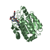
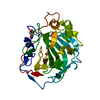
| Title | HaloTag Engineering for Enhanced Fluorogenicity and Kinetics with a Styrylpyridine Dye | ||||||
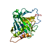
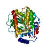
 Components Components | Haloalkane dehalogenase | ||||||
 Keywords Keywords | FLUORESCENT PROTEIN / HaloTag / Fluorescence / Imaging / mammalian cells / fluorescent reporter / channel dye / directed evolution | ||||||
| Function / homology |  Function and homology information Function and homology informationhaloalkane dehalogenase / haloalkane dehalogenase activity / response to toxic substance / membrane Similarity search - Function | ||||||
| Biological species |  Rhodococcus sp. (bacteria) Rhodococcus sp. (bacteria) | ||||||
| Method |  X-RAY DIFFRACTION / SYNCHROTRON / MOLECULAR REPLACEMENT / Resolution: 1.45 Å X-RAY DIFFRACTION / SYNCHROTRON / MOLECULAR REPLACEMENT / Resolution: 1.45 Å | ||||||
 Authors Authors | Stein, A. / Liang, A.D. | ||||||
 Citation Citation | Journal: Chembiochem / Year: 2021 Title: HaloTag Engineering for Enhanced Fluorogenicity and Kinetics with a Styrylpyridium Dye. Authors: Miro-Vinyals, C. / Stein, A. / Fischer, S. / Ward, T.R. / Deliz Liang, A. | ||||||
| History |
|
- Structure visualization
| Structure viewer | Molecule: MolmilJmol/JSmol |
|---|
- Downloads & links
-Download
| PDBx/mmCIF format | 7ond.cif.gz | 146.3 KB | Display | PDBx/mmCIF format |
|---|---|---|---|---|
| PDB format | pdb7ond.ent.gz | 112.2 KB | Display | PDB format |
| PDBx/mmJSON format | 7ond.json.gz | Tree view | PDBx/mmJSON format | |
| Others |  Other downloads Other downloads |
-Validation report
| Arichive directory | https://data.pdbj.org/pub/pdb/validation_reports/on/7ondftp://data.pdbj.org/pub/pdb/validation_reports/on/7ond | HTTPS FTP |
|---|
-Related structure data
| Related structure data | 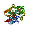 7oo4C 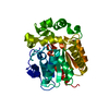 5y2xS S: Starting model for refinement C: citing same article ( |
|---|---|
| Similar structure data |


-Links
 PDBj
PDBj


- Assembly
| Deposited unit | 
| ||||||||
|---|---|---|---|---|---|---|---|---|---|
| 1 | 
| ||||||||
| 2 | 
| ||||||||
| Unit cell |
|
-Components
| #1: Protein | Mass: 34416.242 Da / Num. of mol.: 2 / Mutation: R133C, E143M, F144H, M175Y, V245A Source method: isolated from a genetically manipulated source Source: (gene. exp.) Rhodococcus sp. (bacteria) / Gene: dhaA / Plasmid: pET30 / Production host: #2: Chemical |   Mass: 357.940 Da / Num. of mol.: 2 / Source method: obtained synthetically / Formula: C22H30ClN2 / Feature type: SUBJECT OF INVESTIGATION Mass: 357.940 Da / Num. of mol.: 2 / Source method: obtained synthetically / Formula: C22H30ClN2 / Feature type: SUBJECT OF INVESTIGATION#3: Chemical |   Mass: 35.453 Da / Num. of mol.: 2 / Source method: obtained synthetically / Formula: Cl Mass: 35.453 Da / Num. of mol.: 2 / Source method: obtained synthetically / Formula: Cl#4: Chemical | ChemComp-MG / |   Mass: 24.305 Da / Num. of mol.: 1 / Source method: obtained synthetically / Formula: Mg Mass: 24.305 Da / Num. of mol.: 1 / Source method: obtained synthetically / Formula: Mg#5: Water | ChemComp-HOH / |  Mass: 18.015 Da / Num. of mol.: 617 / Source method: isolated from a natural source / Formula: H2O Mass: 18.015 Da / Num. of mol.: 617 / Source method: isolated from a natural source / Formula: H2OHas ligand of interest | Y | Has protein modification | Y | |
|---|
-Experimental details
-Experiment
| Experiment | Method: X-RAY DIFFRACTION / Number of used crystals: 1 |
|---|
- Sample preparation
| Crystal | Density Matthews: 1.9 Å3/Da / Density % sol: 35.34 % |
|---|---|
| Crystal grow | Temperature: 293 K / Method: vapor diffusion, sitting drop / pH: 5.9 Details: 0.2 M Magnesium Chloride hexahydrate, 20 % w/v PEG 3350 |
-Data collection
| Diffraction | Mean temperature: 100 K / Serial crystal experiment: N |
|---|---|
| Diffraction source | Source: SYNCHROTRON / Site: SLS  / Beamline: X06DA / Wavelength: 1.0000314 Å / Beamline: X06DA / Wavelength: 1.0000314 Å |
| Detector | Type: DECTRIS PILATUS 2M-F / Detector: PIXEL / Date: Mar 20, 2021 |
| Radiation | Protocol: SINGLE WAVELENGTH / Monochromatic (M) / Laue (L): M / Scattering type: x-ray |
| Radiation wavelength | Wavelength: 1.0000314 Å / Relative weight: 1 |
| Reflection | Resolution: 1.45→47.1 Å / Num. obs: 90750 / % possible obs: 99.24 % / Redundancy: 6.7 % / CC1/2: 0.999 / Rmerge(I) obs: 0.06203 / Net I/σ(I): 18.68 |
| Reflection shell | Resolution: 1.45→1.502 Å / Rmerge(I) obs: 0.3734 / Num. unique obs: 8968 / CC1/2: 0.965 / Rpim(I) all: 0.1506 / Rrim(I) all: 0.4031 |
- Processing
| Software |
| ||||||||||||||||||||||||||||||||||||||||||||||||||||||||||||
|---|---|---|---|---|---|---|---|---|---|---|---|---|---|---|---|---|---|---|---|---|---|---|---|---|---|---|---|---|---|---|---|---|---|---|---|---|---|---|---|---|---|---|---|---|---|---|---|---|---|---|---|---|---|---|---|---|---|---|---|---|---|
| Refinement | Method to determine structure: MOLECULAR REPLACEMENT Starting model: 5y2x Resolution: 1.45→47.1 Å / Cor.coef. Fo:Fc: 0.973 / Cor.coef. Fo:Fc free: 0.965 / SU B: 1.106 / SU ML: 0.043 / Cross valid method: THROUGHOUT / σ(F): 0 / ESU R: 0.07 / ESU R Free: 0.069 / Stereochemistry target values: MAXIMUM LIKELIHOOD Details: HYDROGENS HAVE BEEN ADDED IN THE RIDING POSITIONS U VALUES : REFINED INDIVIDUALLY
| ||||||||||||||||||||||||||||||||||||||||||||||||||||||||||||
| Solvent computation | Ion probe radii: 0.8 Å / Shrinkage radii: 0.8 Å / VDW probe radii: 1.2 Å / Solvent model: MASK | ||||||||||||||||||||||||||||||||||||||||||||||||||||||||||||
| Displacement parameters | Biso max: 52.28 Å2 / Biso mean: 11 Å2 / Biso min: 4.48 Å2
| ||||||||||||||||||||||||||||||||||||||||||||||||||||||||||||
| Refinement step | Cycle: final / Resolution: 1.45→47.1 Å
| ||||||||||||||||||||||||||||||||||||||||||||||||||||||||||||
| Refine LS restraints |
| ||||||||||||||||||||||||||||||||||||||||||||||||||||||||||||
| LS refinement shell | Resolution: 1.45→1.488 Å / Rfactor Rfree error: 0 / Total num. of bins used: 20
|
